This week, how immune cells can be caught on camera as they exit blood vessels, a new design of lensless microscope that sees cells in 3D, how sound and heat can be used to find faults in materials and how something as small as an atom can be seen under an electron microscope. Plus, news that nerve transplants can correct metabolic disorders, the World's first fishhook, bionic contact lenses that project emails into your eyes, are statins safe and why are mirror reflections still blurry close up for the shortsighted...
In this episode
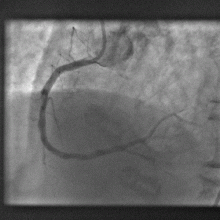
- Statins in the water?
Statins in the water?
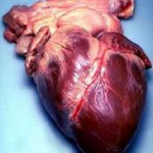
Cholesterol-lowering "statin" drugs are safe and a very effective way to 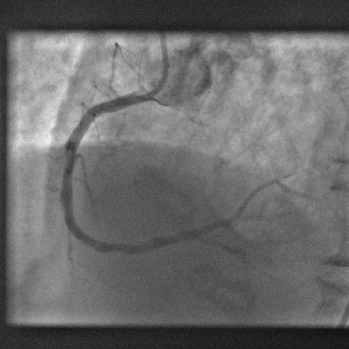 reduce the risk of heart attacks, new research has confirmed.
The latest study, which is published in the Lancet medical journal this week, is a long-term trial coordinated by the Heart Protection Study Collaborative Group.
Over 20,000 patients were recruited into the trial and randomly assigned to receive 40mg of simvastatin, or a placebo, over a 5 year period.
The two groups were then followed up for a further 6 years afterwards to look at mortality and particularly cancer risk. This is because initial trials, despite showing dramatic reductions in heart disease rates and protecting against prostate cancer, suggested that statin therapy might nonetheless raise colon cancer rates by over 50%. This research was, however, not based on a randomised control trial, leading doctors to question the validity of the conclusions.
Consequently the new trial was convened and shows, convincingly, a 23% drop in heart attacks and other vascular disease rates in the statin recipients compared with the controls. The beneficial effect of the treatment also persisted throughout the duration of the follow up period, suggesting that statins carry a long-term legacy benefit that argues for early intervention.
Importantly, comparing the cancer rates between the placebo and statin recipient groups showed, despite numerous cancer diagnoses being made, there were no significant differences between the two groups.
Together, these results suggest that, as Payal Kohli and Christopher Cannon point out in a commentary article also in the Lancet, "prolonged treatment with statins is indeed efficacious, safe, and has long-lasting beneficial effects, even after discontinuation of therapy."
01:20 - The view through a confocal microscope
The view through a confocal microscope
with Dr. Abigail Woodfin, Queen Mary University of London
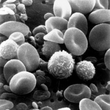
Ben - The optical microscope revolutionised science. It gave us access to an unprecedented world too small to see with the human eye. It's still a relatively simple design, essentially a light source and a series of lenses that magnify the image that you're looking at. There are also some very simple ways that we can adapt it to get increased resolution, a very tight depth of field and even three-dimensional images. Confocal microscopy was invented by renowned scientist Professor Marvin Minsky back in the 1950s and it's now being put to excellent use by, amongst others, Dr. Abigail Woodfin from the Centre for Microvascular Research at Queen Mary University of London. Abigail, first of all, could you just tell us a bit more? What is confocal microscopy?
Abigail - Okay, so confocal microscopy refers to the technique of looking at three-dimensional objects through a series of two-dimensional slices, much like an MRI machine would look through your body during a scan. After you've taken the series of slices down through a three-dimensional object, they're reconstructed to rebuild the 3D object that you're looking at. And that enables you to get much better resolution than if you were looking at the entire object in one go.
Ben - So this is very much like computerised tomography. So for example, when you take a series of x-rays and then put them together, you get that wonderful view that you can sort of flip-book through the image and see an entire 3D aspect rather than just having to look at the one plane, the one single two-dimensional image. How does that actually work?
Abigail - So it's used in conjunction with fluorescence - illumination of the objects you're looking at. So what we do is we use antibodies which are generated to specifically bind one particular protein and the antibodies have a coloured fluorescent tag bound onto them. So when you add these antibodies to your cells or tissues, or whatever you're interested in, they bind to your particular protein, and you then use laser excitation to see the location of these fluorescent antibodies. The objects we're looking at are so small that they are essentially transparent in the absence of using these coloured tags to mark the particular structures.
Ben - What sort of structures are you actually marking out with these tags?
Abigail - We're specifically interested in the blood vessels and how white blood cells or leukocytes react with blood vessel walls during inflammation. So we would use antibodies which bind to proteins within the blood vessel wall such as one called PKAM and we also use cells where a gene for green fluorescent protein from jelly fish has been inserted into the white blood cells so the white blood cells themselves also have a green fluorescent colour.
Ben - So there's lots of tricks you can employ to make sure that you're only seeing the bits that you want to see. Under your microscope or in the images that you get, what does the rest of the surrounding tissue actually look like? Is it completely invisible?
Abigail - It depends. When you're looking with transmitted light or say, a normal white light, which has all of the different wavelengths of light in, then you can see sort of some textures and some structure, you can see blood flowing through blood vessels, and you can see the white blood cells as transparent spheres rolling along the inside of blood vessel walls. However, when you're looking with the laser excitation to look at the fluorescent tags which you've got in your tissue then only the fluorescent tags will be picked up. Everything else will be negative or won't have any colour.
Ben - So, quite often with advanced microscopy techniques, what we need to do is take the sample that we want to look at and then we freeze it or we section it, or we prepare it, we stain it, and ultimately it means that it certainly can't be part of living organism. This sounds like if you're seeing blood flowing through things that you can actually use this with something in situ while it's still alive. And so, rather than having to try and take a moment frozen in time and use that to guess what's happening, you can actually watch the processes taking place inside blood vessels.
Abigail - Yes, that's exactly right. Confocal microscopy has existed for some time, but the time it took to take one of these 3D images was such that it was not really possible to look at dynamic cellular interactions and also, we needed the ability to add the fluorescent tags into living cells. So, the work we've been doing has involved fluorescently tagging the living cells and looking at the interactions with white blood cells with blood vessel walls in living tissues.
Ben - So if in order to see this in 3D, you need to take the series of slices, it's going to limit you for how many essentially frames per second you can take. What is the temporal resolution? Are you seeing this in close to the 24 frames per second that we see on cinema screens or are we looking at something at a little bit more time lapse.
Abigail - It's a little bit more time lapse. For the work we've been doing most recently, I've been taking one of these 3D images every minute. I could increase that slightly to doing say, two per minute, but for the size of tissue structure that I want to look at that's my sort of temporal limit of resolution.
Ben - Still, that's a phenomenal step forward in what we're actually able to see and create videos of these incredible processes happening. What's it actually told us so far about physiology? What's it led us to learn?
Abigail - So the study of the interaction of white blood cells with blood vessel walls has been going on for some years, but has been limited by our ability to really look at these dynamic interactions in real time, in vivo, or on living tissues. So, now that we've managed to get this increased spatial and temporal resolution, we've been able to actually watch the process of white blood cells migrating through blood vessel walls and into the surrounding tissues.
And answer, questions that have not been able to be answered previously using the sort of snapshot images of exactly what route the white blood cells take through the vessel wall, how long they take to do it, do they move in just one particular direction, or do they sort of change directions. And we've managed to identify that the white blood cell migration can exhibit a sort of multidirectional behaviour so that, they go out with the blood vessel wall, they can also in some cases return back into the blood flow.
Ben - So it's obviously opening some doors that were not open before, what do you think will be the next step? How are we going to make this better and take it further?
Abigail - I would say, improving the temporal resolution would enable you to look at faster processes. The things I look at are sufficiently slow cellular interactions that this temporal resolution is sufficient. But faster temporal resolution would enable you to look at faster processes. Higher spatial resolution would enable you to see the smaller structures and interactions.
I think also, increasing the quality of the fluorescent tagging would enable you to see different things. So for example, the protein PKAM in blood vessel walls I mentioned earlier is what we use as our blood vessel marker, and that's the best marker that we have found. But if we could develop ways of fluorescently tagging a whole host of different proteins within the blood vessel wall then that would enable us to deduce things about the functions of those proteins as well.
Ben - And that will give us a whole new multilaminar way of looking at these things, I suppose. Well thank you very much, Abigail. That's Dr. Abigail Woodfin from Queen Mary University of London.
![View with floaters in the eye == {{int:filedesc}} == Impression of floaters against a blue sky. Made by [[User:Acdx]] in Photoshop. == {{int:license}} == {{PD-self}} [[Category:Floaters]]](https://www.thenakedscientists.com/sites/default/files/styles/medium/public/media/Floaters.png?itok=aYiqMWqF)
09:37 - Lensless Microscopes
Lensless Microscopes
with Professor Changhuei Yang, University of California Institute of Technology
Dave - Over the last 400 years or so, microscopes have been developed so much they would often be unrecognisable to 17th century scientists. They can use invisible wavelengths of light, electrons, or even atoms to see with and some are even able to watch individual atoms moving around. But almost all microscopes have one thing in common - the lens. It might be magnetic or electric instead of being made of glass, but there is still an element which does the focusing. However, Professor Changhuei Yang from the University of California Institute of Technology is attempting to do away with the lenses in a microscope altogether.
So Professor Yang, what are the problems with putting a lens in a microscope?
Changhuei - Well, you run into cost issues. Those sophisticated, optical elements in a conventional microscope do cost significant amounts of money to fabricate to implement it well. You also run into the fact that you get astigmatism, chromaticity. Basically, distortion that is intrinsic in a lens that you would have to live with or contend with.
Dave - So essentially, they're just a very complicated, difficult thing to make. If you can get away without them, it just makes the whole thing easier and cheaper?
Changhuei - Exactly and that's basically what the research going on in my group is trying to do, is to basically come up with other ways of doing microscopy, in which lenses are not needed at all.
Dave - Where did you get the idea of doing this from?
Changhuei - Well the idea sort of came from the fact that a lot of us see floaters in our eyes. I'll just describe it to your audience that - floaters are basically debris that float around within your eyeball and when it get close to the retina itself and if you look up into a clear blue sky, the shadow that those floaters will cast on to your retina will be picked up as sharp images.
What is interesting about floaters is that if you have floaters in your eyes, I encourage you to try the following experiment. Try removing or putting your eyeglasses, or just try focusing or defocusing your eyes and you'll notice that those floaters look equally clear no matter what you do. And this says that floaters, the way you see them, doesn't depend on the lenses in your eyes or eyeglasses for doing imaging.
Dave - So these are the little patterns which you see, structures which float around, especially when you look at some bright light.
Ben - I can vouch for this. I have a couple of floaters that I've been ![== {{int:filedesc}} ==</p><p>Impression of floaters against a blue sky. Made by [[User:Acdx]] in Photoshop.</p><p>== {{int:license}} ==</p><p>{{PD-self}}</p><p>[[Category:Floaters]] (c) View with floaters in the eye](/sites/default/files/media/Floaters.png) aware of since I was a very small child and you're exactly right. They always seem to be equally well focused regardless of whether I'm wearing glasses or not wearing glasses, even if I have contact lenses and the floaters are still there and they're still the same. For me, they look almost like bacteria under a microscope appropriately. They look like these sort of rod-shaped blobby things with no real structure.
aware of since I was a very small child and you're exactly right. They always seem to be equally well focused regardless of whether I'm wearing glasses or not wearing glasses, even if I have contact lenses and the floaters are still there and they're still the same. For me, they look almost like bacteria under a microscope appropriately. They look like these sort of rod-shaped blobby things with no real structure.
Changhuei - And the reason why you see them very clearly is because they are very close to your retinal layer itself. So just like if you put your hand very close to a table, you can see a clear shadow image. The same thing happens with these floaters. And by the way, these floaters are fairly tiny objects. They are typically on the order of a hundred microns upwards and yeah, when I see them, I see them with very good details. And this suggests that there is really other ways that you can do microscopy. For example, if you're really interested, you can imagine taking objects that you want to see and inject it directly into your eye and then use this floater phenomena to see things.
Dave - I'm guessing you're not suggesting we do this!
Changhuei - No, of course I'm not advocating that. But the thing is this, thanks to the fact that cell phones now almost always contain a cell phone camera. That actually allows us to have technologies that can do a very cheap imaging using this strategy.
Dave - So you'd essentially just take whatever you're trying to look at and put it directly onto the sensor from a cell phone camera or I guess something which you can just buy off the shelf.
Changhuei - That's right. So they serve as the artificial retina and if we actually take cells and put them on or grow them on those chips, we should be able to actually get some sort of middle resolution imaging performed with it.
Dave - So how good a picture can you get by just taking a cell and putting it onto a camera chip?
Changhuei - Okay, so the typical resolution you can get that way is about 4 microns. To set that in context, a typical cell length is about 10 microns in diameter. So you can't really see features within the cells this way, but you can definitely tell the presence of the cells. And what we did is we further improved the resolution by coming up with an approach in which we take a bunch of snapshots of these cells as they're laying on top of this sensor chip, with the illumination light source being scanned around and that actually gave us enough information that we can then do processing to get microscopy resolution.
Dave - So this is based on the idea that if you sort of say, hold your hand a little bit away from the table, and you move a light around that's going to move the shadow around.
Changhuei - Exactly, and notice that even if the pixel size on the sensor's chip is fairly large, if you take enough of those images where the shadows shift incrementally, you would have collected enough information that you can then later process to get a high resolution image out of that.
Dave - So, if your shadow is half on one pixel and half on the other pixel, if you move the light a bit, it's going to be slightly more than half on the first pixel, and less on the second pixel.
Changhuei - That's right.
Dave - And you can use that information to work out exactly where the shadows should be sitting?
Changhuei - That's right.
Dave - So you essentially got your microscope which is a camera with the light moving around over the top, so would you actually physically move the light around?
Changhuei - The way we implement the light movement is simply have a display in which we have the display showing a round blob of white light and then what that does is that that blob of white light simply moves around on the display and that creates that different angular shift of the illumination that we require for doing imaging.
Dave - Brilliant! So where would you actually see this being used?
Changhuei - So we see this as being useful for both biology and biomedical applications. So for example, in biology, one of our collaborators is a stem cell researcher and when he grows stem cells, and when they starts differentiating, some of those stem cells actually become highly motile.
They move everywhere on the chip and it makes it very difficult for him to actually track where his cells are going. But if you actually grow those cells on this sensor chip and do microscopy level imaging using our approach, you can then automatically trap the sells no matter where they are on the chip itself. So you won't have to actually go and find the cells, you can just take a sponge of snapshots and then later, process it appropriately.
Dave - So it's not just cheaper. It's actually doing something you couldn't do with another kind of microscope.
Changhuei - That's right. So a conventional microscope typically has a very limited field of view. We're talking typically about 100 microns by 100 microns. This new technology allows us to see over the entire area of a sensor chip which is typically on the other 5 millimetres by 5 millimetres.
Dave - I guess and also, it's a lot cheaper so you could use it in a 3rd world kind of situation.
Changhuei - That's right. One of the applications associated with this is that you can actually use this potentially to look at TB cell cultures, TB bacterial cultures. The way it is done right now is you would have to stick this into - the TB bacteria culture into an incubator and then remove it at regular intervals to actually examine it under a microscope system.
If you think about that whole process where it's really old, labour intensive, and you also have to actually run the significant risk of having the samples being contaminated due to this constant shuttling between the microscope and an incubator. With the systems that we have, we can actually simply stick the entire imaging system into an incubator, and have the incubator send out the information either to a wire outside the incubator, or through Wi-Fi, and that allows you to actually image the cell in real time, and track them without actually having to remove them from the incubator.
18:14 - Nerve transplants wire themselves into host brains
Nerve transplants wire themselves into host brains
Embryonic nerve cells transplanted into a recipient brain survive, 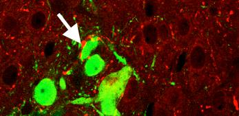 wire themselves up and can even correct a metabolic disorder in vulnerable individuals, scientists have discovered.
Successful brain repair in the future will almost certainly depend upon the implantation, within the injured or diseased nervous system, of healthy cells that can replace those damaged by degeneration.
But the fate of these fresh cells, when added to an abnormal brain, and whether they can functionally wire themselves up to and support existing nerve circuits, is poorly understood.
Now scientists have proved that these cells can do this by using cell transplantation techniques to remedy an obesity-triggering metabolic disorder in mice.
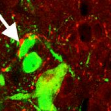
Writing in Science, Harvard scientist Jeffrey Macklis and his colleagues transplanted into the obese mice, which lack the receptor in their brains for a satiety-signal called leptin, 15,000 nerve cells collected from 13 day old mouse embryos.
The cells, which also synthesised a glowing-green pigment to discriminate them from the hosts' brain cells, were implanted into a region of the brain known as the medial hypothalamus, which controls appetite.
Five months later, the researchers looked for signs that the donor neurones were still present in the brain and were speaking electrically with their host brain neighbours.
The implanted cells, they found, had wired themselves in and were functional. They also found that, compared with control animals and mice that had received grafts from other brain regions, the treated animals were 30% lighter and had more normal blood glucose measurements.
This, say the scientists, is "proof of concept for cell mediated repair of a neuronal circuit controlling a complex phenotype."
20:37 - The coldest point water can remain a liquid has been calculated
The coldest point water can remain a liquid has been calculated
If you ask any group of school children what temperature does pure water freeze at and you will normally get the answer 0 degrees celsius, which is the standard answer and it is the temperature below which ice is more stable than water, but that isn't the whole story.
It is quite easy to get liquid water below this temperature by just putting a bottle of very clean water in a freezer, because although a large ice crystal is more stable than liquid water, a very small one isn't so most of them shrink and melt before they get big enough to be stable, but how cold can you get liquid water?
Valeria Molinero and Emily Moore from the University of Utah decided to do away with experiments and try and solve the problem in a computer. This isn't easy, as forming ice intrinsically involves a lot of water molecules, and to get a meaningful result they had to model over 30 thousand water molecules interacting with one another, and they had to model water molecules as a single lump rather than as three atoms..
They found that as the water cooled, more and more of the water formed tetrahederal structures which were somewhere between ice and liquid water, in both structure and density which they call intermediate ice. This can then either convert to ice proper, or into a disordered glasslike structure when it finally froze.
After all this work they finally found the theoretical lowest temperature you can cool water to without freezing is -55 celsius.
This might seem quite academic, but supercooled water is very important in many types of cloud, and has been discovered there at -40 celcius, and understanding how water behaves at these temperatures will help understand clouds and so assist with weather and climate predictions.
22:52 - First seafaring fisherman
First seafaring fisherman
The world's oldest tackle, together with evidence of deep-sea 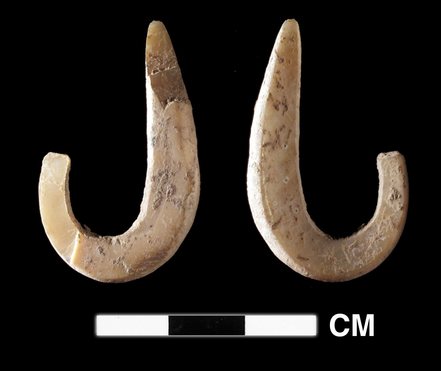 fishing 40,000 years ago, has been unearthed in East Timor.
Early human migrants, including those who first set foot in Australia 50,000 years ago, were clearly competent mariners. The journey from Eurasia into the northern part of the Australian continent would have involved crossing a 1500km wide deep-water archipelago, negotiatable only by boat and good seamanship. But archaeological evidence of these seafaring feats is sparse.
Now, however, Australian National University scientist Sue O'Connor and her colleagues, working at a site in East Timor called Jerimalai, have discovered evidence that early human inhabitants of the coastline were catching and consuming fish from the open ocean as far back as 42,000 years ago.
Bones belonging to over 20 different fish species were recovered from an excavation at the site, 50% of them belonging to so-called pelagic (open water) dwellers like tuna. To catch these, the Jerimalai settlers would have to have ventured out to sea armed, most probably, with nets.
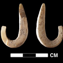
But even more exciting is the discovery, also amongst the finds, of two primitive fishhooks carved from pieces of trochus shell and dating from about 24,000 years ago. This is the earliest evidence of fishhook manufacture yet described.
As the team point out in their description of the findings, published this week in Science, "Capturing fish such as tuna requires high levels of planning and complex maritime technology. The evidence implies that the inhabitants were fishing in the deep sea."
25:15 - A Diode for light
A Diode for light
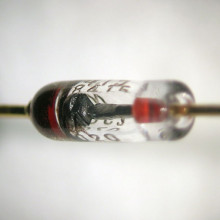
For many years engineers have wanted to use light instead of electricity to build circuits with. It moves slightly faster, but more importantly signals can pass one another without interfering. This lack of interference makes it very difficult to make light beams interact when you want them to.
One of the simplest electronic devices is the diode, effectively a one way 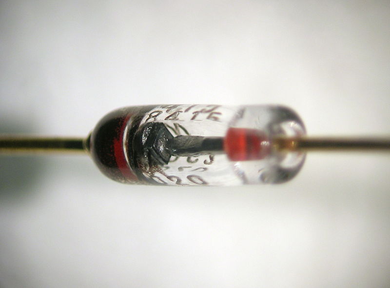 valve for electricity, in light they would be a true one way mirror, and are very difficult to build. So far they have been mm or even cm across and it hasn't been possible to integrate them into a silicon chip. But Caroline Ross and collegues at MIT have managed to do this.
They have built a structure which involves a tiny conducting silicon loop electrical resonator which can absorb energy from light which is passing through a nearby piece of garnet covering half of the loop.
This garnet is magnetic as well as transparent, so applying a magnetic field means that the loop will absorb a different colour of light in one direction to the other, so if you pick your colour it will be absorbed 100 times more in one direction but than the other.
In the first case this type of device would probably be built in front of a laser to stop reflections interfering in its operation, but a diode is a vital component for building more complex photonic circuits in the future. Which amongst other things should help route the optical signals which move most of the data around the internet.
27:40 - The Safety of Statins
The Safety of Statins
with Richard Bulbulia, University of Oxford
Ben - Also this week, a study published in the Lancet has confirmed the safety of the widely used class of drugs known as statins. These are the most commonly used drugs worldwide for heart disease and are taken by millions of people globally. A randomised heart protection study back in the mid-1990s found the drugs to be highly effective against heart disease but subsequent epidemiological studies did raise some fears of an increased risk of cancer associated with taking them. Now Richard Bulbulia from the University of Oxford has followed up the participants in that study from the '90s to have a look at any long term effects. Richard, thank you ever so much for joining us.
Richard - Good evening, Ben.
Ben - What were they really looking at back in the '90s? Were they just confirming that statins did work and did do what we think they should?
Richard - Observational studies made it clear, 30 or 40 years ago, that people with higher levels of bad cholesterol had increased risk of vascular disease and to test whether this association was causal, people did large, randomised trials such as the Heart Protection Study which lowered cholesterol and consequently lowered vascular risk by around one quarter. But the same epidemiological studies that highlighted the relationship between cholesterol and vascular risk also showed an increased risk of certain cancers and other causes of non-vascular death with lower cholesterol levels, and those of us who sort of responsibly interpreted that data suggested that these findings were due to something called reverse causality whereby it's the disease such as the cancer which causes the low cholesterol rather than the converse. But the concerns were out there and they substantially delayed the widespread use of statins.
Now, the Heart Protection Study which reported its main results in 2001 had reassurance in that there were no excess risks of cancer or other non-vascular deaths associated with taking simvastatin 40 mg for around 5 years. But that 5-year period was really too short to reliably address the prevalent concerns about the risks and safety of lowering cholesterol in many millions of people. And for that reason, we carried on following up all 17,000 surviving Heart Protection Study participants for a further 6 years.
Ben - So what have you actually been doing to follow them up or are you just looking at medical records and incidences of different diseases or are you continuing to investigate more deeply what the lifestyle factors are? 
Richard - We followed up the survivors in two ways. We asked them to complete postal questionnaires and the vast majority of the participants did so, and on these postal questionnaires, they told us certain things about their statin use after the trial period, whether or not they've been to a hospital, had any clinical events, and also indicated whether or not they'd be happy to receive a subsequent questionnaire the following year. That questionnaire procedure was augmented by accessing national registries for cancer incidents and they're death certification for people who either had cancers or died during the follow up period.
Ben - Surely, after the original trial, the people who had been taking placebo, as it was a controlled trial, they must've then been offered the statins. So surely, actually, the conditions have changed. How do you take account for that?
Richard - That's a very important point. At the end of the trial, it was clear that everybody in the Heart Protection Study would benefit from at least discussing whether or not they should take the statin with their GP and all were encouraged to do so. Gratifyingly, over the 6-year period, more and more people in both treatment groups have began taking the statin therapy, so by the end of the trial, the average use of statins was around 75% in both original treatment groups. So, our long term follow up results, which were in the Lancet this week, actually assessed the effect of the initial 5-year randomisation to either simvastatin or placebo over an 11-year period.
Ben - I guess the fact that people did start taking them after, also means that you could actually stratify your results and show that people have been taking it for this long, you see the following effects, but if people have been taking it for twice as long, then either you see more effects or you still don't see any effect which should help you to be able to say the concerns about cancer were in fact not actually applicable.
Richard - That's correct and there were three main findings in our study. The first two connected are looking at benefits. I mean, it's important to remember that statins are incredibly effective form of treatment. During the randomised phase of the trial, the absolute benefits of statin therapy increased as treatment continued. With year on year reductions of around one quarter, after the first in trial year, and second, the absolute benefits that those originally allocated simvastatin accrued during the in trial period persisted in the post trial period. That is to say, the people who were originally on placebo and then switched to statin therapy after the trial had closed, never caught up with the original simvastatin group. And those two findings do show that starting statins early and continuing them long term is necessary to maximise the reductions in major vascular events.
And following on from that clear message, it's also very reassuring to note that over an 11-year period, there was no suggestion of an emergence of hazard on cancer either globally or in certain specific subtypes of cancer or indeed other forms of non-vascular disease and specifically non-vascular death emerging in this large cohort of trial participants.
33:18 - Bionic Lenses, Night Vision and Stress on the Brain...
Bionic Lenses, Night Vision and Stress on the Brain...
with Babak Parviz, University of Washington; Erno Hermans, Donders Institute; Zhengwei Pan, University of Georgia; Andrew Shuiteman, Kew Gardens
Flashing emails before your eyes
A
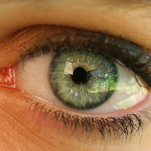
contact lens capable of displaying electronic information, quite literally in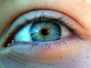 front of your eyes, is being developed by US scientists.
Challenges include powering the device and displaying complex information such as text, But Babak Parviz and colleagues at the University of Washington have so far combined a miniature radio receiver and light source within a lens and wirelessly sent single pixels of data safely into the eyes of rabbits.
Babak - You can imagine your cell phone might some information to your contact lens and the contact lens has an antenna that can receive the information, has a radio that can process the information, and drawn a display with it, and there are some extra focusing mechanism that allows you to see the image. If you think about our daily routines we interact with a number of displays, the TV screen and the computer displays, the cell phone display this, but in a sense, we don't really need all of them, if we have a personal display that is our contact lens, we can get rid of all these extra displays and just have one display that is personalised to the user.
---
Stress on the Brain
Sudden stress can cause
changes in connections between different regions of the brain
Short bursts of stress are known to sharpen senses, impair abilities to deliberate and create fearful arousal, although how this was achieved by the brain wasn't known.
Now, by exposing human volunteers to clips of violent and non violent films and imaging their brain activity, Erno Hermans and colleagues from the Donders Institute have discovered the changes in the brain causing these responses.
Erno - We see change in the way brain regions communicate with each other. Those are regions that are involved in reorienting attention and also regulation of your autonomic nervous system, and of regulation of the stress system. So what we see here is that those regions sort of become active together and form a network as if they're integrating information across all these domains. This might be a model for studying what happens in potentially traumatic situations.
---
Non-Stop Vision in the Dark
A new
materialthat can glow for over two weeks after just minutes of exposure to sunlight has been developed by scientists at the University of Georgia.
A mixture of chromium ions embedded in a matrix of zinc, gallium and germanium oxides can soak up the energy in visible light, releasing near infra-red wavelengths and providing night vision for up to 15 days even in cloudy or overcast conditions.
Zhengwei - The first one lies in military defence and the law enforcement, and the other thing is solar energy absorption and storage. The third application is we can make the material into nanoparticles so that we can out these particles this molecule and particle into the body so that it can link to some tumour cells for bio-imaging.
---
Night Flowering Orchid
The first night-flowering orchid has been discovered by scientists at Kew Gardens.
The flower, now named Bulbophillum nocturnum, originates from the Island of New Britain near Papua New Guinea, and was found to open a few hours after dusk and remain that way until a few hours after dawn. But it only does this for one night.
The species is the only orchid known to flower only at night, with the reasons behind this behaviour a subject of speculation for Orchid expert Andre Schuiteman from Kew Gardens.
Andre - Out of 25,000 species of orchids known approximately, this is the first one, officially we are certain that it is flowering at night. This is quite strange because related species flower during the day time. We think the flower opens at night because it is pollinated by flies that are active after dark or maybe early in the morning, when it's just getting dawn, probably to escape predators.
More information on the orchid can be found on the Kew gardens website at
kew.org.

37:21 - Alien Hikers - Planet Earth Online
Alien Hikers - Planet Earth Online
with Professor James Bullock, Wallingford’s Centre for Ecology and Hydrology
Dave - Alien, or non-native species, can have serious effects on the landscape and indigenous wildlife. Recent research in Australia has found that hikers could be partly to blame...During just one season in a national park there, hikers were found to carry up to two million plant seeds just on their socks!
James Bullock, from Wallingford's Centre for Ecology and Hydrology, was one of the authors of the study, which was based on some of his earlier work in Dorset, Planet Earth podcast presenter Sue Nelson joined him at a picturesque section of the River Thames in Oxfordshire to see what they could find....
James - Riverbanks are actually one of the most invaded habitats we have in Britain, so the sort of species you might see along here are Himalayan Balsam or Japanese Knot Weed, all of which cause problems with riverbank stability affecting native biodiversity. What you will see also this time of year is the chestnut leaf miner, a lot of chestnut trees their leaves will be removed by mining of this moth caterpillar. Actually, we can see over here, just next to us, are Canada geese which have been established in this country for a long time and cause a lot of problems, not just affecting native bird species but by pooing in waterways cause a fertilisation effect which can effect what's growing in the water as well.
Sue - So the effects then of invasive species are quite wide and varied, it's not just a threat to biodiversity in some cases?
James - No, they have quite a wide range of effects. They can effect biodiversity directly, but they can also affect aspects of the natural world which are of more direct impact on humans, such as bank erosion, pollution of waterways, causing problems with grazing lands, so you have invasive species spreading across grazing areas which affects the ability of animals to graze on them.
Sue - Now the Australian study found that different parts of a hiker's clothing could spread different numbers and types of seeds.
James - There are some seeds which have hairs or bristles on them which are probably evolved to allow their dispersal by animals; we come along with our socks or our trousers, almost like an animal skin and it forms a nice material for these seeds to attach to. So, the more woolly the clothing you're wearing and the more bristly the seed the more seeds will get dispersed by people.
Sue - How did this come out of research from Dorset?
James - We were interested in quite a different aspect of dispersal in Dorset. We were working on a species called Wild Cabbage there, which is quite a rare species restricted to our coastline and it forms quite discreet populations along the coastline which have been sitting there for probably hundreds of years. So, we were interested in the reverse side of the question - what limits this species to grow where it grows and what are the possibilities for its dispersal?
Along the coastline of Britain we have loads and loads of footpaths and one possibility we thought of was that hikers could take the seeds of the cabbage around. So we did an experiment where we found that seeds rather than socks or trousers would stick into the mud on hikers boots and could be transported very long distances, the natural dispersal by wind could take seeds a maximum of about 200 metres, but hikers could take the seeds over five kilometres.
Sue - Is that why you want to know? Is that why you do this sort of research in order to predict possibly, or can you even predict when you've got millions of seeds capable of sticking to somebody's hiking socks what species are going to be transported where?
James - Yes. The prediction is the key here. What we want to understand for alien species or non-native species is what species is transported, how far they're transported and what are the mechanisms of their transportation. And the reason we do that is not simply understanding, but then we can use this sort of information in models of spread of these species to work out how we might limit that spread, so a lot of work on alien species so far has been saying, ok, we find out where the populations are growing and we go and try and kill them in some way.
A much more efficient and effective method would be to prevent the movement of these species in the first place and so that's what we're working towards - understanding the whole process of spread so we can look at the crunch points and limit that spread by especially targeting dispersal.
Sue - How would you advise hikers, how on earth are they going to limit the spread of seeds?
James - Yes, that is a big question and it could sound a bit over the top telling people to be very careful about what they're transporting. But certainly in the study in Australia, this is a highly protected area which is under threat from these European plant species coming in, so in that case, the recommendation has been to put signs up for walkers, to educate walkers, to say before you walk into these remote areas just clean your trousers and socks off, just pick the seeds off before you spread them into these pristine areas. So, I think in very specific circumstances, where we're trying to protect particular areas, there is something that can be done.
42:39 - Imaging with heat
Imaging with heat
with Tony Dunhill, Rolls Royce and British Institute of Nondestructive testing
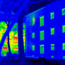
Tony - Thermography is the use of temperature differences of a part and it involves using a thermal camera. Thermal cameras have come on in this world greatly over the last 5 or 7 years. We're now able to use various forms of heat application. The most traditional is passive thermography where you just look at a part and look for the hotspots and that's used a lot in power stations, engineering structures where you might see a failing at a point and it gets hot just before it fails. Active thermography, you apply some heat and that can be done with a flash lamp with electrical induction or it can be done with a laser. So the usual method is to apply some sort of heat, look at how that heat dissipates with time, and then within that time period, there'll be a point where a defect will reveal itself.
Ben - Heating components, especially metals, tends to lead them to expand. Are you risking sort of obscuring problems that you might see with other techniques by perhaps expanding the material and therefore, filling up cracks that might be there?
Tony - Well, when we say we heat it, we heat it to a minute level. I mean, we're talking about changes of temperature at a maximum of about 2 degrees. So just by picking the thing up, you're putting the same amount of heat in as we're using. So, the change in profile of the defect due to the heating is going to be minuscule. Although I have to say, if the crack is under a lot of compressive stress, it is quite easy for heat to transfer itself across the crack and so that, often is a poor detector under those circumstances whereas if the crack is open, then the technique is much more viable method.
Ben - So how else can we apply the heat? You said there'd be several different methods beyond literally just flashing a very bright hot bulb at it. How else can we make these cracks show themselves?
Tony - There's two methods that are being worked at the moment. One is induction thermography where you put the parts in an induction coil and that gives you a local region that you're heating and if it's a long part, you can then just pass the piece through. The second method is with a laser spot and this is very useful if you can't get near the part. So like nuclear storage drums or something like that then you can shine the laser at some distance away, you can use a thermal camera to see the heat spot caused by the laser, and then you can scan the laser around, and where it comes across a crack then you get a change in the dimensions of the heat spot.
Ben - So this isn't just something that has to be done on an engineer's bench somewhere. You can actually use thermography in situ.
Tony - Yes, as long as you can get a line of sight to the place you're interested in and they are beginning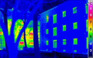 to be quite useful boroscopes that can take infrared light. So, the access issues have become less of a problem and distance can be quite effective. You can use the laser technique over quite a few meters and still have a fairly sensitive technique. You could even go through various forms of glass, provided it'll transmit the infrared.
Ben - So we have lasers, we have induced heating, are there any other means that we can create the heat and see these cracks?
Tony - Well quite often, you don't necessarily need heat, but you could use cold. You can pass cold air on the rear surface of the part and it'll suck the heat away and where there's a defect in the way that might retain heat. As long as you get this heat differential across the defect, then that's usually what you're looking for.
Ben - New coatings are being developed all the time and new alloys which change the way that the surface will oxidise. Does this mean that you're constantly having to review exactly how you apply these techniques in the light of these new developments?
Tony - Where we have new materials, then we have to understand how that material property will affect the technique we're applying. So yes, there's a sort of baseline exercise that you have to do. But I mean, most metals behave in a similar way and most ceramics behave in a fairly similar way, so you can make a judgement. And we have good theoretical understandings from our research centres that support us and this thermography one in particular at Bath University where we take advice from them as to the different effects that new materials would have on the techniques we apply.
Ben - That was Tony Dunhill from Rolls Royce.
48:08 - Imaging with sound
Imaging with sound
with Professor Bruce Drinkwater, Bristol University
Bruce - Well the ultrasonic waves, similar form to acoustic waves, so we know they're travelling in air, but they also travel in liquids and solids. And so, dealing with bits of metal then ultrasonic waves travel really very nicely in those bits of metal. So, we have a series of sources which emit ultrasound into the metal structure and they're reflected from all of the interior structure which obviously includes cracks. We can then pickup with our microphones, ultrasonic microphones, actually they're small electric elements but doesn't really matter there. We can pick up that reflected energy, process it and from that, produce an image of the interior of the component.
Ben - A lot of the materials that we might be looking at to try and find cracks are actually very highly, very precisely made materials in the first place. Often, they can be a single metallic crystal. Does that actually affect the way that the ultrasound would travel through?
Bruce - Yes. The crystal structure is utterly paramount to how waves travel in so that the single crystals are an isotropic in their material properties for which as far as ultrasound is concerned means that waves travelling in different directions go at different speeds, and you can imagine that if we're not very careful, that could create all sorts of confusions we have to compensate and take account of the fact that the velocities are different and the speeds of sounds are different in different directions. And if we do that then we can produce images as if the velocity was uniform.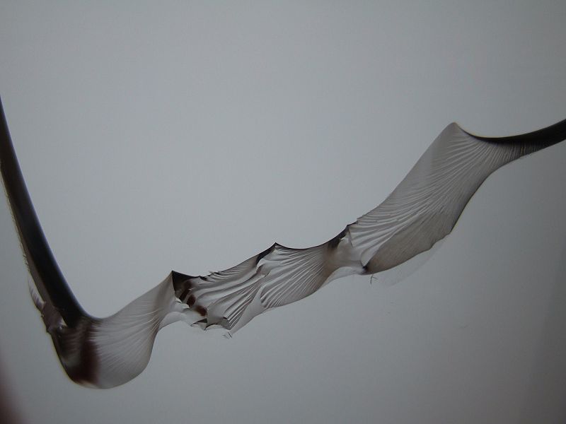
Ben - So essentially, you use some kind of algorithm or some kind of computer modelling to compensate for how you know the sound would travel in a pristine example and that therefore means that you can compare your example to pristine, and easily see if there's something wrong.
Bruce - Yes, we need to have a model of the structure we're trying to test in order to correctly image it, and one of the big challenges is when you go to a component and try to make a measurement where there's uncertainty in that model. And so, one of the things we have been working on is what we call autofocus techniques where your camera automatically adjusts to put things in focus whereas we have been working on ways of trying to automatically extract this picture of what the structure is like. So this model without knowing it beforehand or say, it's the particular principle orientation direction is unknown. But we can extract that from the data itself if we know something about the structure like, for example, how thick it is.
In terms of detecting cracks, the real challenge is, somewhat different to the medical challenge where you've got this very rich image. The interior of the piece of metal is you'd have thought relatively simple and it is, but there's a lot of scattering from other things within the material like grain boundaries, although they don't occur in the single crystal example that you just talked about but in most metals, there are many grain boundaries, all reflecting ultrasound and there may or may not be cracks and so, the challenge is to achieve that high resolution imaging performance of the crack surrounded by these other scatterers which basically set the signal to noise ratio of your measurement or the best possible measurement that you can make.
Ben - And I assume that signal to noise is ultimately what will limit the resolution with which we can view this and the actual minimum size at which we can first detect that crack.
Bruce - Yes, and that's always the challenge, to detect the smallest possible crack and that's where we're always trying to push the limits of what's possible there and that's what I'm interested in as a researcher.
What does imaging atoms really show us?
Dave - Pretty much all of the kinds of microscopy which can see atoms are forms of electron microscopy. Some of them are actually atomic microscopy way or either firing electrons at a surface and getting them to bounce off or you're firing atoms at a surface and getting them to bounce off and you can draw a picture from that but actually, the more common one is actually called scanning electron microscopy.
These have a very, very sharp point and they just scan this point across the surface on an atomic resolution so you build up lots of lines. And the point is moved up and down to either produce a constant force which is called atomic force microscopy or a constant electric current which is called scanning tunnelling electron microscopy. The point moves along and it goes up and down to get this constant current. So what you're seeing is something to do with the electrons on the surface, either it's how hard they're pushing the point up and down or it's to do with how well they conduct and how well electricity can move up into the point. By changing the voltage, you can actually get different pictures and you can look at electrons at different parts of the atom and you can actually do some very cunning bits of science on that.
Ben - So although we're looking at different properties of the atom itself, what we build up with is an image built by inference. We're inferring that because of the change in electrical properties or the change in mechanical properties, there is therefore something there and it's on such a small scale that we can actually infer the exact position of an atom.
Dave - Yes, that's right. And something which is very neat which has done a while ago which I saw in the news was that they were doing things on a sheet of graphene which is such a regular repeating structure that you can subtract away this background pattern which you're getting in your picture, and just leaving the atom or molecule on the surface, so you could actually see hydrogen atoms attached to carbon atoms which is absolutely minute and you couldn't possibly see it in other ways.
54:32 - Why couldn't I focus on a reflection when close to the mirror?
Why couldn't I focus on a reflection when close to the mirror?
So even though the TV ought to be close enough, why does the mirror keep it blurry? We posed this question to Dr Brian Robertson, research associate at the photonics and sensors group in the Department of Engineering, University of Cambridge...
Brian - In answer to your question, you have to focus a camera or your eyes because as your light leaves an object it spreads out but to get a good image, the lenses in your eye have to bend all the light from one point in the object onto one point on the retina in the back of your eye.
If you're close to an object, light is spreading out more quickly so the light needs more bending to produce a sharp image and come into focus than if you're far away. The eye adjusts to these distances by changing the shape of the lens - short and long sightedness occurs when the eye is less able to accommodate these changes.
Light reflecting from a mirror gives the impression that the object is behind the mirror and you get this impression because the light having reflected from the mirror is moving exactly as if it were coming from an object behind the mirror. This is not just true of the direction of the light is travelling, but it's also how the light is spreading out.
Your eye does exactly the same job focusing on an object that appears 10 metres away in the mirror as focusing on the actual object 10 metres away.
So, if you're short sighted, the object, in this case the television screen, still appears blurred regardless of how close you are to the mirror.
Diana - On the forum, RD said that it doesn't matter how close you get to the mirror when an object is a certain distance from it. If you're short sighted, then the lens in your eye cannot adjust for the amount of spread the light has taken over that distance. He also mentioned that a convex mirror can cause the object to appear even farther away as it spreads the light even more. Again, making it blurry if you're short sighted.
Next week, nails on a blackboard...







Comments
Add a comment