Miniature lungs, breasts and other organs are being grown in dishes so scientists can study how they form, why they succumb to disease and how toxins, drugs and poisons affect them. Organ models like these are rapidly replacing animals for many lab experiments. But are the days of the petri dish also numbered, as computer models, like the "virtual physiological human", become more powerful. We talk to scientists using and developing all three. Plus, a new coating stops joint replacements loosening, magnetic therapy for strokes, and plants do long division...
In this episode
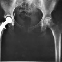
01:03 - Coating firmly anchors bone implants
Coating firmly anchors bone implants
A new coating technique that makes bone bond tightly to an artificial joint has been 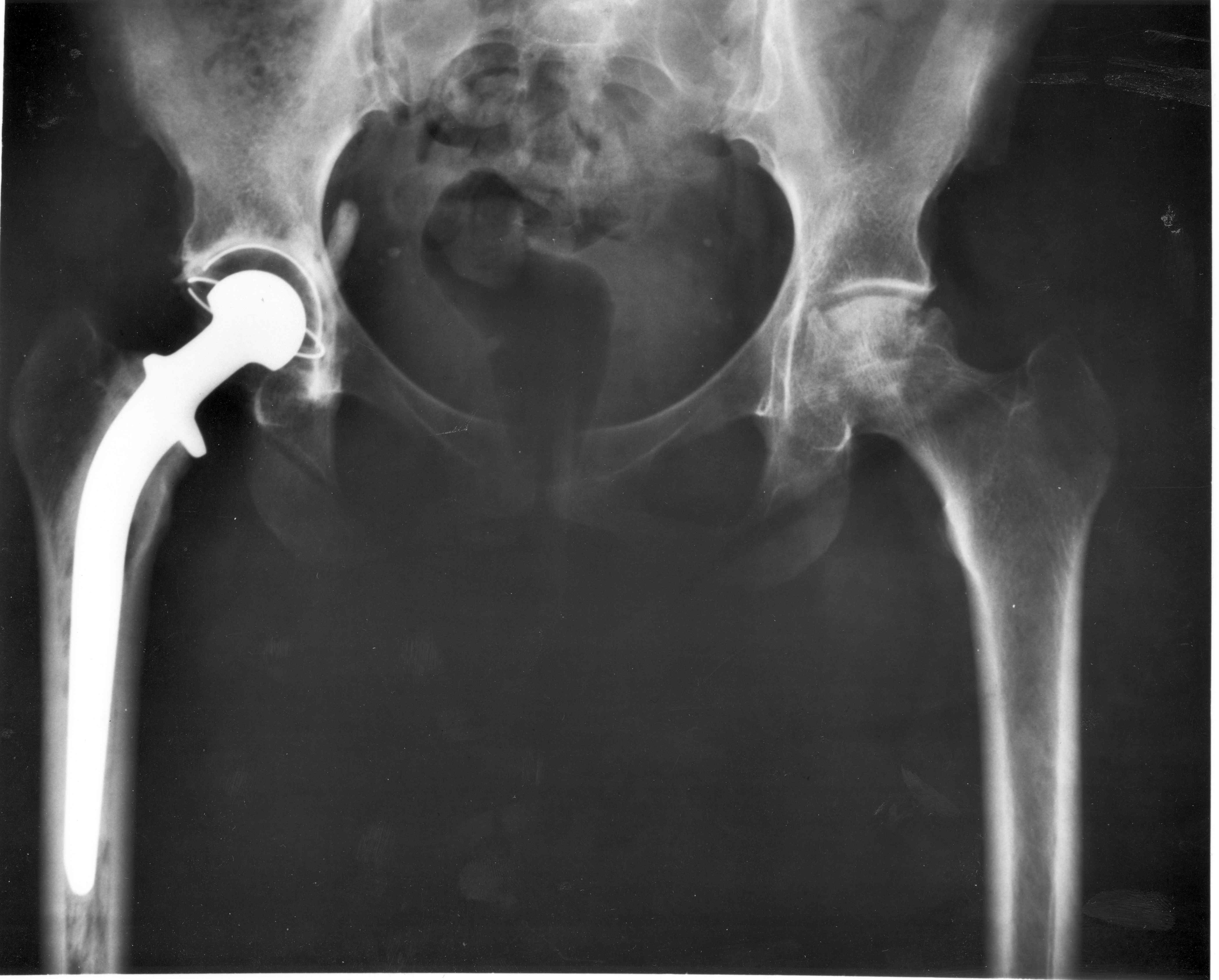 developed by scientists in the US.
Millions of joint replacements are performed each year, but in under a decade at least 12% of them will require surgical revision, half of these because the prosthesis has worked loose.
Currently, replacement joints are anchored in place using a bone cement compound called poly-methyl methacrylate (PMMA), which is disadvantageous for several reasons: most importantly, it has a different stiffness to native bone, so when the bone bends or flexes it moves a different amount to the cement, causing cracking and encouraging the two to part company.
Also, as PMMA is setting, it releases a large amount of heat, which can damage nearby tissue and also makes it difficult to include within the cement chemicals or growth factors that might help to promote local healing, or bone repair.
Efforts were made to produce porous prostheses intended to encourage bone to grow into the device, locking it in place, but these too have largely been abandoned owing to poor bone in-growth.
Now, step forward MIT's Paula Hammond. She and her team have instead developed a technique to apply a multi-layed, growth-factor impregnated coating to the surfaces of titanium or plastic prosthetic devices.
The base layer of the coating, which is about 1/5000th of a millimetre thick, contains hydroxyapatite (HAP), the very same calcium phosphate compound found naturally in bone.
Atop this HAP layer is a series dissolvable layers made from the bio-friendly polymers poly-beta-amino ester and polyacrylic acid. Impregnated into each of these layers is a growth signal called bone morphogenetic protein-2 (BMP2), which strongly stimulates the growth of new bone-building cells known as osteoblasts.
The thickness of these dissolvable layers, which build up like an onion skin and are formed by repeatedly dipping the prosthesis into a solution of the chemicals and then allowing it to dry, can be varied according to need. For a thicker layer, dip more; for a thinner layer, dip it fewer times.
Implanted into the shin bones of experimental animals, prostheses treated with the new coating showed steady release of the BMP-2 growth factor into the surrounding bone over about a 30 day period, and an ensuing in-growth of bone-building osteoblast cells.
Examining the implants under the microscope at time points up to 18 months later showed that the dissolved top layers had disappeared and new bone had grown up to and knitted itself into the HAP (hydroxyapatite) layer. There was no evidence of any tissue reaction to the implant.
Even more convincing was that, compared with untreated control implants, the force needed to remove from the bones prosthetics treated with the new coatings was over 30 times greater, and the test implants frequently came away with pieces of newly-grown bone still attached.
Writing in Science Translational Medicine where the work is published, the authors nonetheless acknowledge that they have worked only with rats so far and the technology needs to be tested in larger species and more realistic clinical settings.
But with the world's population ageing as it is, the demand for improved prosthetics with extended lifetimes has never been greater...
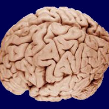
04:36 - Can magnets help stroke patients?
Can magnets help stroke patients?
In the UK one person has a stroke around every five minutes. This potentially  debilitating condition occurs when blood vessels in our brain either have a blockage or a haemorrhage, which stops oxygen reaching parts of our brain. While the areas affected change from case to case, around 20 to 30 percent of patients experitence a condition called aphasia which affects our ability to read, write, speak, or understand language. This condition can be particularly frustrating as patients find them suddenly unable to communicate with with their family, or their needs and wishes to hospital staff.
In patients with aphasia, two areas of the brain are likely to be affected, (which in 97% of people are in the left hemisphere of the brain). These are Broca's Area, in your frontal lobe which affects how we produce speech and Wernicke's Area, in the cerebral cortex, which affects how we understand speech. In order to minimise the impact of stroke, doctors aim for fast intervention, to restore the oxygen supply to deprived areas as quickly as possible. However, once damage has been done the current treatment only involves Speech and Language Therapy which, in the first few weeks aims to provide practical help for patients to communicate their needs on a daily basis.
This week, however, a paper has just come out from a team at the McGill University in Montreal who have been trying to improve language function using transcranial magnetic stimulation. This involved using a magnetic coil which when you put it next to someone's skull, creates a changing magnetic field inciting an electric current in the nerve cells just on the other side of their skull. It's at a really low intensity, but if you held it over the motor areas in your brain you'd feel muscle twitching as your nerve cells were activated.
They hoped that by using this stimulation on areas deprived of oxygen during a stroke, they'd be exercising these areas and it might help bring those back into use a bit quicker. They tested 24 patients, giving half of them real stimulation and half of them fake for 20 minutes a day, along with 45 minutes of speech therapy for 10 days. They found that those who received the real transcranial magnetic stimulation had a 3 times greater recovery rate, as measured on aphasia language tests than those who didn't. Obviously, it's a small study with only 24 patients, so they want to test this method on much trials, but it's looking very positive.

07:42 - Buying a pint with bitcoins
Buying a pint with bitcoins
The virtual currency bitcoin has long been used by gamers to buy extra features online. But now users can spend their savings on real life pints as in the past few weeks some British pubs in London and Cambridge have started to accept the online coins in the real world. Here's the Quickfire Science on the currency with Hannah Critchlow and Dominic Ford.
· Bitcoin is an online currency which can be exchanged from person to person without the need for a bank or other financial intermediary.
· It can be used in any country and aims to be independent of any central control.
· The currency was first described in 2008 in a paper by an anonymous person or group under the name Satoshi Nakamoto.
· A year later the same group published the first freely available bitcoin software and issued the first pieces of currency
· Each bitcoin is a specific piece of computer code. As well as exchanging bitcoins, users can 'mine' for new currency with their computers.
· Computers 'mine' by solving complex mathematical equations which hold bitcoins in the network - the difficulty of these changes depending on how many coins are being released.
· There are plans to limit the number of bitcoins to 21 million which are predicted not to be uncovered until the year 2140.
· Although there is no central bank, all transactions made using bitcoin are noted in a public transaction log. This means no-one can try to pay for multiple purchases using the same coins.
· Each transaction on the log is also 'signed' by a private code that is associated with the users' bitcoin wallet - proving it came from the correct owner.
· To use bitcoins in the real world the shop produces a QR code for the purchase which the customer then scans with their phone using a bitcoin app.
09:50 - Keeping an eye on diabetes
Keeping an eye on diabetes
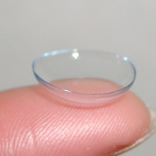
Diabetics could one day be able to monitor their blood sugar levels using bionic contact lenses, instead of having to resort to painful finger pricks. Researchers have developed a fuel cell that runs on tears, which they say could power lens-mounted glucose sensors.
The idea of using lenses for diabetic monitoring has been around for years, as 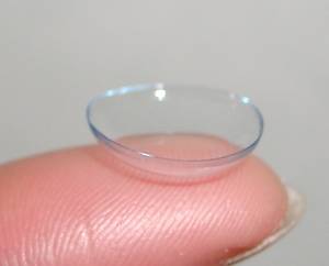 glucose levels in tears track blood glucose levels. An electrical glucose sensor in a lens would be in constant contact with tear fluid, and could produce readings for an on-the-spot display that could be easily read by the wearer. But such a sensor would need a power source, and so far this has proven a major stumbling block.
An international team led by Sergey Shleev at Malmo University, Sweden, may have come up with a solution by developing a biofuel cell that runs on the ascorbate (vitamin C) and oxygen naturally present in tears. The cell uses two organic catalysts at the anode to oxidise ascorbate, and the enzyme bilirubin oxidase to reduce oxygen at the cathode. Together, the electrodes power the cell.
Using human tear samples, the team showed the fuel cell could generate power from tears without altering their glucose content.The cell's power output is very low - theoretical calculations estimated it could produce up to 22.1µW in an actual contact lens, based on the ascorbate concentrations and rate of tear flow in a human eye. But the team say this could be enough to power a tiny sensor, particularly if the electrode sizes could be increased by using both sides of the lens.
'An ascorbate/oxygen biofuel cell could be a suitable power source for glucose-sensing contact lenses,' the team summarise in their paper, adding that all the components used appear to be biocompatible.
13:17 - Can imagination affect what you see?
Can imagination affect what you see?
Does our imagination affect what we see and hear?
Our perception, what we understand about what's going on in the world around us, is a combination of all of our senses. What we see and what we hear comes together to tell us what's going on. But, if our senses disagree with each other, we tend to come up with the wrong answer.
One famous multisensory illusion, called the McGurk illusion, shows that if we  hear someone saying the sound /ba/, but we see them saying /ga/, we think they're saying /da/. So, when our senses are disagree we amalgamate them and believe the position in the middle. This week researchers at the Karolinska Institute have published a paper in Current Biology that asked, 'What about our imagination?' If we imagine hearing something, could it affect what we see and play into these multisensory illusions?.
They recreated three famous perception experiments with one element imagined rather than seen or heard to see if they still work under these conditions. One experiment showed a cross of ramps going past each other with a disc at the top of each ramp. As the discs ran down the ramps the participants were asked to imagine a sound happening, some at the point where the discs passed each other, some before and some afterwards. Those that imagined the sound happening just as those discs passed each other thought that they saw the discs bouncing back off each other - the illusion still worked, even though the sound was imagined.
Similarly, they re-did the McGurk illusion. They asked participants to imagine the sound /ba/, showed them a video someone saying /ga/ and they still perceived /da/. These results show that multi-sensory illusions are still effective even if of those senses is imagined rather than actually happening. Researchers have suggested that this implies neural networks which register perceived and imagined senses have some cross over, so what we imagine is included in our perception of the world. They hope a greater understanding of this process will be able to contribute to treatment of some psychiatric disorders, like Schizophrenia, where people are unable to distinguish between what is real and what is imagined.
15:46 - Maths maketh mortgage success
Maths maketh mortgage success
Did homeowners with a poor command of maths cause the global financial crisis, 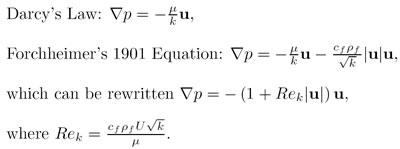 US researchers are wondering?
Kindled in the US subprime mortgage market, the ensuing economic shockwaves ricocheted around the globe, dragging most of the industrialised world into the most severe recession since the Great Depression.
Most of the blame has thus far been levelled at "irresponsible bankers" who, critics argue, shouldn't have lent money to mortgage buyers who could not afford the debts. But to what extent are the borrowers themselves to blame, and what factors might influence this? And could poor numerical ability be the cause?
To find out, University of Lausanne researcher Lorenz Goette and his colleagues analysed the mortgage records of subprime borrowers dating from 2006-2007.
The individuals concerned were contacted and, over the telephone, underwent basic tests of their numerical and verbal abilities. These results were then compared with the likelihood that these same individuals had defaulted on their mortgages and for how long.
What emerged was a very strong relationship between mortgage default duration and weak mathematical ability. Those least adept at adding up spent, on average, twice as long in default compared with those scoring the highest marks.
This relationship held even after controlling for factors such as verbal ability, meaning that it wasn't just a borrower's inability to understand the terms of a mortgage that was responsible; instead it appears that numerical ability drives behaviour downstream of securing a mortgage and this is the critical factor.
The researchers conclude their paper in PNAS by highlighting two policy implications of their findings: First, the complexity of mortgage products, making them hard to comprehend, has been blamed for contributing to home repossessions. But altering just this, the team say, based on their results, won't solve the problem.
Instead, they highlight second that improved financial education of homeowners is what is required, suggesting that a follow up "randomised control study", offering financial training to some homeowners but not others and comparing the outcomes could test the importance of this intervention.

18:15 - Breastfeed for a higher social class
Breastfeed for a higher social class
Being breastfed could help you climb the social ladder, a new first-of-a-kind study has shown. 
Breastfeeding is known to confer a range of health benefits, but it might also contribute to being upwardly mobile.
UCL epidemiologist Amanda Sacker and her colleagues compared the social classes of the parents of two large groups, each of more than 16,000 people, one born in 1958 and the other in 1970, with the social classes of the individuals themselves when they were aged 33, and asked whether they had been breastfed as infants.
Compared with their parental social class, breastfed participants in either of the two age groups were 25% more likely to have climbed the social ladder and 20% less likely to have been downwardly socially mobile by age 33.
The effect, which the researchers suggest is causal based on their results, was also related to how long a baby received breast milk, with the strongest relationship emerging for those babies who were breastfed for longer than four weeks.
The team speculate that the effects may be both nutritional, reflecting the essential fatty acids, antibodies and growth factors supplied by breast milk, and emotional, reflecting enhanced mother-baby bonding and the fact that breast-fed babies may be less susceptible to the effects of stress.
But, these are just speculations that require formal investigation. Writing in Archives of Disease in Childhood, the team point out that "more research is needed on the association between between breast feeding and child cognitive and socio-emotional development to elucidate the causal mechanisms through which breast feeding can have lifelong implications for health and well-being."
It should also be emphasised that the effect is probably not retrospective either...
26:21 - Modelling! Medical mimicry
Modelling! Medical mimicry
with Kelly BeruBe, Cardiff University
Modelling. Not the catwalk variety, but medical mimicry!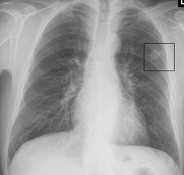
When researchers want to peer inside the human body to understand it better, one option is to create an artificial model of the area they're interested in: be it lung, heart, or breast. They can then tweak this artificial system to test drugs and see how it reacts, in order to understand it better. We were joined by Kelly BeruBe, a researcher at Cardiff University, who's doing just this for lung tissue.
Chris - So, what's involved in trying to make a model lung in a dish?
Kelly - A lot of work, that's for sure and a lot of innovation, and a lot of patience. But first, I should begin by probably telling you what a model is. With modelling, I would say it's basically like being a hobbyist. You reproduce an item of interest. So, in my case, we're looking at building a replica of the airway region of the lungs and that's the conducting part of the lungs, the big tubes where air moves in and out because that's the area where, when you inhale something, it takes the biggest hit. So, we like to focus on that region for any kind of inhalation studies. For using models, well, they're useful because you can replicate them endlessly, very quickly, usually, very economically whereas if you compare that with animals, they're very expensive. You'd have to let them live their whole life span if you wanted to make a comparison with the human situation. The other thing is that, with using models that you create, you can then change little parameters on those models and then measure those. That gets rid of the problem with extrapolating animal data to the human situation because if you're using human tissues like we use, we use lung cells and tissues donated from patients then you have human endpoint data.
Chris - So, you make a number of compelling positives for why this is a good idea, but it's presumably not trivial to make something that behaves, looks, and functions as a lung in a dish.
Kelly - Yes. I mean, we've been working on this now for 10 years and we used to work with animals for the past 15 years and weren't getting anywhere. And then I think about 2003, we were able to buy human tissues, you could procure cells from human tissue banks. We started dabbling with them and all of a sudden, questions that were eluding us for years, we were getting the answer to those very quickly in a matter of a year or so. So, we decided that we were going to leave the animals and move right into human tissues and it was like a Lego system. We just started playing with different cells, different media, different bioreactors because we needed to make the cells in 3D to work. If they're in 2D like in a petri dish, you don't get the same reactions that you would get if it was in the human body.
Chris - How do you get the cells to grow into that 3-dimensional structure that the lung is (first point) and second point, if you look in a lung, they're not just one type of cell. There are many different types of cell, aren't there? There are muscle cells, glandular tissue, epithelial cells that line the airways, and then special surfactant making cells that make the air sacs where the respiratory exchange takes place. It's really complicated.
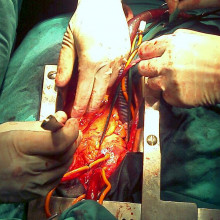
Kelly - Chris, you could work in my lab. You sound like you know a lot, but yes, there's over 40 different cell types in the lung and they're divided into 3 different regions. You have the upper respiratory system which traps things and tries to prevent them from getting into the lower lung, then you have the thoracic region where we work in that has a lot of defence mechanisms. Then in the lower lung, the distal lung, you have the alveolar region where you breathe, where people exchange oxygen and CO2. You don't want anything in that area because you'll get inflammation. So, we work in the thorax where it has the highest number of defence cells in that area because its job is to stop things from getting into where you actually breathe. Now, in that region, there's about 7 key cells and what we do is we take the basal stem cells from donors and the cool thing here is that we can use medical waste tissues. So, if you have an operation and they open you up, and they have to nick out some tissue, and they throw it away, we can buy that. They usually incinerate that so we buy it, we take out the stem cells or the cells that we're interested in and then we regrow them in bioreactors. These are special membranes in, they look like little petri dishes, like little cups about the size of a pea and the top part is open to the air, and the bottom part you feed. That's just like how we breathe. When we breathe in air, it goes over the tissue and you'll get your nutrients from below.
Chris - Do the cells know where to go?
Kelly - Yes. This is like a military secret. I could tell you, but then I have to kill you type thing because it's all patented technology, but I'm sure the other people will tell you this. Using the 3D culture media that we created, it has the right amount of hormones and chemicals that tell the cells when and what to turn into basically, at what time and it grows a multilayer into 7 different cell types and you get your mucus secretion, you get your cilia doing the samba, beating back and forth. It looks just the piece of tissue that you would take out of a person.
Chris - And what sorts of questions can you ask and answer with this that you couldn't do previously when you were working with say, mice or other rodents, other experimental animals?
Kelly - We know we can accurately dose ourselves because when we used to do installation work, where you inject a fluid with a particulate matter or something into the lung. We were never really sure where the compounds were going. We would just put it in and faithfully think we've put a milligram in and we're hoping it's dispersing throughout the whole lung, but you'll never know. So, this way, we can accurately put a dose on that's environmentally relevant. So, we're not overdosing the cells.
Chris - And it's very reproducible and very consistent. So I suppose, getting the numbers up to a way that you can say this is statistically valid is easier.
Kelly - Absolutely. We buy in about 500,000 cells from donors and we'll get about 400 pea-sized lungs to work on and they last for about 2 weeks which is rare, so you can do acute, chronic, and repeat toxicity testing. In terms of cosmetics, this is a big deal now because in March this year, the EU had this directive where they ban the sales of any cosmetics that were tested in animals. So, it's often an alternative device now to industry where they've always relied on animals.
Chris - And you can do that testing for them.
Kelly - That's the whole idea now. We're trying to develop a model where people can test compounds that are going to be used for cosmetics and beauty purposes as well as obviously safety testing for medicines. Think of all the stuff that's in the air - air fresheners, aerosols, pesticides, perfumes, make up. It's an endless list of things that we're inhaling into our lungs.
Chris - The fact is that my lung is a part of me and I have an immune system. A petri dish doesn't. it doesn't have a blood flow. There are other aspects of the model that you can't reproduce with your dish. So, how do you know that you're not missing something, even though you're using human cells?
Kelly - Very good question. The thing is that people have to realise is that when you're working with alternatives, it's a reductionist type model and the whole idea is that if it's not complex, you can avoid a lot of confounding factors. So, you can tweeze out little delicate things that you would miss. For example, the immune system or if it's a female and the hormones are in the way, those reactions could mask things that you're trying to find. So, what these alternative methods do is they give you a look at the least complex situation and something of interest should stick out. You can build up and use more complex models to try to answer your question such as using in silico or other in vitro models of different tissues in the body.
34:08 - A 3D breast in a dish?
A 3D breast in a dish?
with Dr Jonathan Campbell, Cambridge University
Jonathan, firstly, why are we so interested in modelling breast tissue?
Jonathan - Well, it's the most prevalent cancer in females. So, if you are able to model the normal situation, you are going a long way towards turning on oncogenes and introducing the tumour microenvironment into that normal situation, and actually, studying physiologically what happens in oncogenesis.
Kate - So, by modelling a breast tissue as it normally is, we can introduce 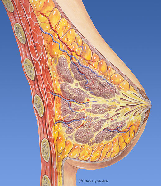 cancerous genes and cells and work out what happens and hopefully, from that point I suppose, work out drugs to try and stop that process?
Jonathan - That's right.
Kate - So, are you using human cells like Kelly is?
Jonathan - Well at the moment, we've produced a model that I'd say is physiological, to the point where it even produces milk. But we are now moving into producing a human model of breast tissue.
Kate - So, what's that model at the moment you're using instead of human tissue?
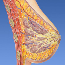
Jonathan - So, what we have is, a natural biomaterial which is effectively collagen and also proteoglycans. We introduce the principal components of a mammary gland into that model. First, the fat cells and also, the breast tissue itself which is the branching epithelium and we are effectively redeveloping the model in the dish in a natural form.
So, in the breast during puberty and in pregnancy, you get this massive elaboration of this tree and we're actually effectively doing that again in the dish. So, we're quite confident it's physiological.
Kate - You're putting these fat cells together, but you said it even lactates. Are there certain cells to do that or once you've put it in a model, does it automatically work out how to do that and form a breast tissue as a whole?
Jonathan - There's a certain amount of it automatically doing that, yes. You give them a little bit of nurturing and they'll seem to do what they do best.
Kate - Those fat cells that you're putting, do they come from humans or do they come from another sort of test animal?
Jonathan - As it stands, it's purely cell lines and these are derived from mouse.
Kate - How similar is breast tissue in mice and humans because if they're forming the structure that they used to, that they know about like you just mentioned, is that sort of physiological structure and layers, are they similar in both mice and humans?
Jonathan - They're relatively similar and obviously, the gland is a lot bigger as we know in the human. The compartment of different cells is slightly different. So, in a human, there are more fibroblast for instance.
Kate - Kelly just talked about having a pea-sized lung. If you're taking mice breast tissue, how small is that within our dish?
Jonathan - The scaffold that we use is actually, you cut it to any size. In terms of these small organoids formed within the scaffold, we're talking of about half a millimetre in length.
Kate - You're working towards building a model that replicates human that you can then introduce these cancer genes into. Once you get to that point, what can a 3D cell model tell us about breast cancer that other cancer research into genes or within sort of patients who have the disease can't tell us? What's it going to tell us that's different?
Jonathan - What's quite exciting is that we can actually move into looking at primary cells and these forms similar structures, but they form what's known as a basement membrane. So, they're enveloped by a particular array of proteins. What's exciting is that cancer is dangerous of course when it spreads and one of the things they have to do is they have to force themselves away or through this protein mix. So, we can actually monitor that in real time. You can't do that in any other way, so we're able to actually - because it's in the dish, you can actually see what's going on.
Kate - So, you can watch the cancer cells spreading along. Once we know more about that mechanism, what can we use that information for? Can we test new cancer drugs and see how that affects it?
Jonathan - Certainly, we can. The thing to realise about cancer, it's a highly individual disease. So, if we can form these small little mammary glands effectively in the dish and we can move into a human model, then we can test all manner of compounds on individual tissue from individual donors and that's very exciting because each person's cancer is different.
Kate - We're going to be talking in just a moment to some computer modellers who do their modelling theoretically. What advantages do you think that cell modelling has that if you looked at this from a computer point of view, you wouldn't be able to find out?
Jonathan - Computer modelling of course has its place, but the biological situation is much more unreliable and I think it was summed up by a talk I went to actually last week in London by a Professor of Oxford, Professor Phillip Maini who stated that in mathematics, if you divide 1 by 2, you get a half and in biology, if you divide 1 by 2, you get 2. It's basically unpredictable.
Kate - Them's fighting words! We'll see what Katherine and Peter have to say about that in a moment. Thank you very much to Jonathan Campbell from Cambridge University.
39:04 - Computational coding to crack disease!
Computational coding to crack disease!
with Katherine Fletcher, Oxford University Peter Kohl from Imperial College London.
We've heard how we can grow tissue in the dish in order to study biological processes. But what about taking that information, and creating a computer model?
We're joined now by Katherine Fletcher from Oxford University and Peter Kohl from Imperial College London who are doing precisely this. Hello to both of you.
Katherine - Hello.
Peter - Good evening.
Kate - We'll be talking to Peter in just a moment about his use of cardiac 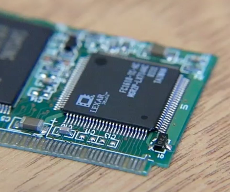 computer models, but first, Katherine, how do computer models compare with delving into the dish?
Katherine - Well, if you're asking me whether a model on a screen is better than a model on a dish, I have to say that neither is as good as you would like them to be. Tissue isn't a whole organism and neither is a computational model. The trick is to make them a useful tool to answer a particular question and where computers are especially good is at integrating lots of different types of information.
You could imagine at a patient level. In the case of breast cancer, you might look at an MRI scan, mammogram results, information about genetic markers, and other blood tests. Maybe the patient's history and risk factors, and if you were able to plug that all into a computer, it would help you sort through the data to find the most interesting points.
At a more basic science level, we could use computers to integrate different readings from experiments by mapping for example the behaviour that's known about cells onto a geometric mesh representing the anatomy of the organism, to for example, find out what is driving a particular type of irregular behaviour.
I'm here on behalf of the Virtual Physiological Human Project whose aim is to make computational models of the human body in health and disease so that different models of different parts of the body can be clumped together to answer different questions.
Chris - Could you just introduce us to the concept of what the Virtual Physiological Human is and how this came about?
Katherine - Yeah. It's a European commission funded, very ambitious initiative using computers to integrate data, going from the sort of subcellular or smaller scale levelled up, as well as going back again from the whole human level, back down and mapping them together in ways that are interchangeable. Since about 2008, the EC has funded 46 projects on a variety of different topics from tumour growth to drug safety, to osteoporosis, and aneurism, each of them working in its own area, but trying to make sure they all use standards so that those models could be used together in different ways in the future.
Chris - So, they speak a sort of common computer language so that if you're developing a model of the kidney, it will talk to ultimately, the model of the heart, so data from one could inform results to another?
Katherine - Absolutely! Doing that in practice is quite tricky. I mean, just getting software to run on your own computer sometimes, you'll run into difficulties, but that is absolutely the idea. By using common computer software languages for example, allows you to then plug them together.
Chris - So, most of the initial setup of the project must've been defining those sorts of standards that the individual groups will work to?
Katherine - Out of the 46 projects that have been funded, one of them was called the network of excellence whose job was to basically serve as a rallying point for researchers to understand what others in the field are doing, and together, develop best practice in standards. And so, there have been a lot of technical things developed through that such as ontology's with the standardised vocabulary for describing a model. If you're interested in this sort of thing, vph-portal.eu is the VPH portal website where you can find all of these information online if you're interested.
Kate - Why can't we create an entire human model that can work all these out at the same time?
Katherine - That's actually technically quite complicated to do that. If you're trying to run a model for example of a human heart beating using a very up-to-date computational model, you have to get that to run not only accurately, but you'd have to get it to run faster than real time because it's no good to predict that a person is going to die of a heart condition in 5 years' time if it takes you 7 years to predict that. So, you have to get these models to be both accurate and extremely quick before they're useful.
Kate - I presume when we start the model, we have to plug in all the data we know to get it to work accurately. Where do we get that data in the first place?
Katherine - There might be information pulled from scientific literatures, so you might find experiments in papers and you can borrow the data from them. It might be coming from real world sources so patients. So for example, MRI data or stands of various body parts that might be diseased. And the trick is finding ways that you can combine them so that you might be able to map the picture of the MRI of a particular person's tumour state to what it already known also about how other tumours of that type grow, and combining them in a meaningful way to get what treatment this patient might respond to best.
Chris - So, where are you at now? So, you have the standards defined, you've got these projects funded to start building models of individual systems. Where are we in terms of actually having a model person in a computer?
Katherine - The idea is still not actually to ever run them all as one individual, at least not as a model stand. The idea is to select the pieces that you're interested in. So, we have lots of very well developed models for example in the cardiac field, musculoskeletal models are also quite good. There's still a lot of interesting things done with viruses and also with cancer. You can imagine all those pieces could all interplay quite well for example.
Kate - And Peter, you're working on one of those projects to do with that cardiac modelling. What do you need to look at in order to model how the heart works on a computer?
Peter - We need to first perhaps start off with agreeing on what we understand then as the terminology of a model. I think the definition that the dictionaries offer, a simplified representation of reality is very helpful because if you appreciate that any model that you might want to use in whatever circumstance is simplified representation of that reality. We also realised that any model regardless of whether it is a lung in a dish or a model of the heart will have its own limitations. If we appreciate that then from there, it follows that the idea and that is coming back to a question you asked slightly earlier, of an all inclusive model of the human is a bit of a contradiction in terms.
Chris - Are we not really in a position where all that the model knows is all that we know? So, until we've done the biology, how can we have a computer that's actually any better?
Peter - If our models did indeed contain all the knowledge we have, that would be great and I don't think we can claim that for any of our model systems including computer models. If they did have that knowledge however, they would be suitable and useful in different ways. One would be to treat them as an expert system that can be interrogated their real use for computational model in such as of the heart is to use them to try and understand better the experimental or clinical data that we receive.
A single cell of the heart is a very complex entity. These cells are governed by processes at the molecular level that are so complex and that interact via multiple feed forward and feedback mechanisms that certainly my brain is not good enough to interpret findings that we may observe in experimental setting. So, running computer simulations at the same level and in parallel to experimental studies can be incredibly helpful in understanding what you're doing. The next step beyond that and that is, I think where it gets really exciting and really interesting is to run simulations first before you go into the lab or before you consider what you might want to do with the patient because you explore the parameter space in theoretical investigations that might allow you to predict what the most likely paths for success might be. Success in this context depends entirely on the application - that maybe development of a drug or a need for an experimental series that gives you a clear-cut answer.
Kate - So Peter, when you've been modelling the heart on a computer, what have you found so far by testing that model?
Peter - One angle that I'm taking is that often experimental researcher who tries to combine experimental and theoretical investigations and these models have been incredibly helpful in planning better experiments. In particular then, we find that our models do not reproduce what we expect. That may sound slightly counterintuitive, but if you have the best conceived model there is and it doesn't quite reproduce what you see, then it highlights a shortcoming in our understanding. It poses a question and science really is all about the question.
Now in terms of more applied aspects such as development of drugs, I wish we had Gary Mirams here with us who is conducting cardiac single-cell modelling at Oxford University and what Gary has done is to look into how one can try and predict side effects of pharmaceutical agents very early in the development stage. You see, the most frequent cause for drugs that may otherwise be very useful that requires them to be withdrawn from the market are cardiac side effects. Now, Gary Miram has developed a technique that is - one might say, reductionist in that he looks at the shape of that electrical signal in a cardiac cell before people look at one single-ion channel type and predict at what might happen. Now, he uses three ion channel types and that may sound really small, but the impressive thing is that his ability to predict the likelihood of cardiac side effect has improved multiple times over what was possibly before. It shows that a simplified model has its place to play in the process of explaining phenomena.
Kate - Thank you so much to Katherine Fletcher from Oxford University and Peter Kohl from Imperial College London.
How does training affect free will?
Hannah - First up, what is free will and how is it involved in trained movement? Dr Tristan Bekinschtein at Cambridge University has this to say.
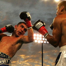
Tristan - Free will is the idea that you can decide something, feeling that you have the ownership of that decision. When you're learning for example to ride a bike or when you're learning to punch someone when you're training to be a serious boxer. Those decisions initially, you have to think about them. They slowly become automatic. So, in a way, you're losing the free will while these things become automatic. To take a decision and reflect on that decision to be sure that you're going to do it and then do it, it takes forever in terms of cognitive processing. It takes 300 or 400, 500 milliseconds With training, the movements become so automatic that you forgot that you were making the decision to move. In fact, you're not making the decision to move consciously anymore and if you're not making the decision consciously, therefore, you're not doing it in a free will manner. Hannah - So, as we learn new movements, we exert free will to control our bodies, but with training, this movement becomes automatic and conscious control is lost. Movement is controlled in motor regions in circuits in the brain, but what happens here as we age? Professor Patrick Haggard from University College London explains.
Patrick - So, I think what happens to the boxer as they become older is that the circuits that allow them to land the punch don't operate quite as fast as they originally did. They begin to slow down just like a lot of our brain function slows down and after a while, they're operating sufficiently slowly that the boxer's conscious experience can actually keep up with them, so he's aware of what he's doing. So, his action control has slowed down over time to the same kind of rather slow speed that conscious cognition operates at and at that point, it's too slow to beat his opponent and he ought to stop.
Hannah - Is this type of phenomena found elsewhere other than during say, riding my bicycle or sports training like boxing? Over to Dr. Gabriel Krieman at Harvard Medical School.
Gabriel - This phenomena is also found in trained musicians playing complex pieces. In many of these situations, consciousness seems to interfere with complex action patterns. What consciousness and free bias is flexibility perhaps at the expense of lower reaction times. Reflexes are faster, but they lack adaptability.
As the question suggests, extensive training can transfer conscious actions into non-conscious reactions. Crick and Kohr referred to this non-conscious reactions as zombie modes.
Both systems, non-conscious reactions and conscious actions are important and have probably conveyed evolutionary advantages.
Hannah - Thanks, Tristan, Patrick and Gabriel.
- Previous How do solar sails work?
- Next Punching into free will




Comments
Add a comment