Scientists have developed a way to introduce colour into body scans, potentially ushering in a powerful new method to detect disease.
 Writing in this week's Nature, Maryland-based NIH researcher Gary Zabow and his colleagues describe how they have developed a family of tiny tracer particles that emit radio signals at different frequencies that MRI (magnetic resonance imaging) scanners can see. The scanners then translate these different frequencies into colours on the images that are shown to the operators.
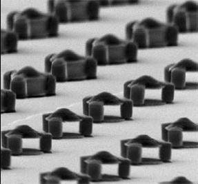
The particles, which range from one-thousandth to one hundredth of a millimetre, resemble nanoscale dumbells consisting of two gold-coated nickel discs joined by a cross member which spans a cavity between them. The nickel particles line up with the scanner's magnetic field, but also produce their own magnetic field running in the opposite direction. As a result, water molecules sitting in the cavities between the nickel discs can be selectively excited by pulses of radio waves at certain frequencies. The particles then produce their own radio waves, which the scanner can pick up and build into a colour image.
The researchers also suggest that the particles could be "functionalised" so that they aggregate preferentially in certain tissues, or remain stable only in certain conditions which means that body scans of the future won't just be shades of grey but technicolour and far more informative!


Comments
Add a comment