Neuroimaging
This week we delve deep into the secrets of the brain. We'll find out how MRIs could be used to read your mind, and how they could help unlock what is going on in the brain of a person suffering from delusions or hallucinations. In the news we'll hear that the process of nerve repair could offer clues to cancer spread, and how it was gorillas that originally gave us malaria. Plus in Naked Engineering, Dave and Meera explore the amazing world of superconductors...
In this episode

- Can you get hallucinations of smell and touch?
Can you get hallucinations of smell and touch?
We put this question to Professor Paul Fletcher...
Paul - You certainly can - you can get hallucinations in any sensory modality. You can get hallucinations of taste, smell, touch, as well as visual and auditory. In fact, in certain cases of mental illness, hallucinations of smell can be exceedingly unpleasant and people can have the belief that they're actually rotting from the inside.
Chris - That's not very pleasant.
Paul - It sounds horrible.

- Why do I talk out loud when I dream?
Why do I talk out loud when I dream?
My own view would be that the nervous system suppresses the motor system when we actually go to sleep. This is why we don't end up acting out more of our dreams, because of this region in the brain stem that stops the motor pathways going through. That will include some of the more complicated movements that make vocalisations happen, but you're still, in your dream, going to be talking and things, aren't you? So, that's why you would talk out loud sometimes - I guess there'd be a breakthrough of that suppression activity.

- Do we really only use 10% of our brains or is that a myth?
Do we really only use 10% of our brains or is that a myth?
We put this to Professor Jack Gallant and Professor Paul Fletcher...
Jack - I think that's definitely a myth. We use a lot of our brains a lot of the time, and it switches back and forth all the time. Which sub-systems of the brain are being used depends on the task and what you're trying to do, but the brain is there for a reason and you're using a lot of it most of the time.
Paul - I was just thinking, the person who started that rumour was probably using 10% of their brain at the time, I think.
Chris - Very good! The point is, if you look at someone who's had a stroke and they may have only lost a small amount of their brain, they nonetheless don't look normal or they may not behave entirely normally, indicating that you need all of your brain. It's just slightly less active at certain times.
Paul - Yes. I mean, one could add that people who've had a hemispherectomy - a whole half of their brain removed at an early age - actually go on to achieve great things intellectually. So, if it happens early enough and the brain is sufficiently plastic, then actually you can do without a lot of the volume of the brain, but I think you would use what was there 100%.

- How does an MRI scan "see" a hallucination?
How does an MRI scan "see" a hallucination?
We put this to Professor Paul Fletcher and Professor Jack Gallant...
Paul - I don't know if that's a great metaphysical question. I mean, the brain scanner is looking at how the brain behaves when it is seeing something that isn't there, so it's not so much interested or able to see the content of that although as we've just heard from Jack actually, the possibility of seeing what the brain thinks is there is possibly something for the future.
Chris - Any comment on that Jack?
Jack - Yeah. I think there's growing evidence that when you have a visual hallucination, what's actually happening is the visual areas of your brain are being activated, essentially top down from inside out and the visual experiences you have in the visual hallucination are -since the brain subsystems were being operated in those cases are visual, then you both experience visual events and you could decode visual events because you're decoding from the same parts of the brain that are encoding visual information normally.
Chris - And hence, what you've got is this system where you think it's real because it's the same bit of the brain that would say, "Yup, I'm experiencing something" but it's just being internally generated.
Jack - Right.

- Are MRI scans safe?
Are MRI scans safe?
We put this to Professor Jack Gallant...
Jack - As far as anyone knows, there is no long term danger from MRI. Magnetic Resonance Imaging involves putting someone in a very large stationary magnetic field. As far as anyone knows, it has no influence on any systems of the body or any biological systems.

01:42 - Nerve repair provides clue to cancer spread
Nerve repair provides clue to cancer spread
Cancer is a disease caused by cells multiplying out of control. It's been known by many names through history - some of them unrepeatable on air, from people and families who've been affected by the disease. One ancient name is "the wound that does not heal", and today, researchers are uncovering many similarities between the controlled cell production required for wound healing, and how this process is hijacked in cancer.
Now new results from a Cancer Research UK-funded team led by Professor Alison Lloyd at University College London have found an important link between nerve repair and how tumours may spread within the nervous system, and they've just published their findings in the journal Cell.
Our nerves are actually pretty good at repairing themselves, although it doesn't always work in the case of severe damage or crushing. For example, nerves can grow back through deep cuts, and it's possible to reattach amputated fingers, toes and even limbs, and have some regrowth of the nerves. And this is all down to special cells called Schwann cells.
Schwann cells act a bit like insulation around nerves - like the plastic coating around electrical wires. Normally, they help the speed up nerve signals, but we know that they're also really important for directing repair.
When a nerve is cut, Schwann cells start growing out into the wound, and start making "guide tracks" for the nerves to grow along. And here's where the new research from Professor lloyd and her team comes in, as they've uncovered some of the complex molecular signals that control the process.
The researchers were looking at exactly how the Schwann cells are controlled and directed into these "guide tracks" by fibroblasts - repair cells that gather at the site of a wound. The team discovered that fibroblasts produced a 'signal' molecule called ephrin B, which is received by 'receptor' proteins on the surface of the Schwann cells called EphB2, standing for Ephrin receptor B2.
It's this ephrin signalling that tells the Schwann cells to organise themselves into tracks, as directed by the fibroblasts, so the nerves can regrow, a bit like traffic police using special hand signals to direct cars into different queues on a road.
And, importantly, the scientists found that switching off ephrin signalling meant that Schwann cells couldn't repair nerve damage, proving that it's a fundamental part of the process. So these findings tell us something important about nerve repair, which will be useful for researchers working on techniques such as nerve grafts, which could repair damaged nerves after accidents or surgery.
So how is this linked to cancer? Well, we know that some types of cancer can spread along nerve cells - and the way they do this looks very similar to the way that the Schwann cells and fibroblasts move as they repair damaged nerves, and it's likely that the same ephrin signalling is involved. 
Professor Lloyd thinks that cancer cells may be acting a bit like an unhealed wound, hijacking the signals that normally repair nerves. In a normal situation, the regrowth signals would be switched off when the nerves have grown across the wound. But in cancer, it may be that the signals don't get switched off and the cancer cells carry on spreading along the nerves, rather than ever settling down and 'healing'.
So understanding how this signalling goes wrong, and how we can block it, could prove to be an interesting lead for future treatments that stop cancer from spreading.
05:35 - New insights into damage done in MS
New insights into damage done in MS
Scientists in Germany have uncovered a previously overlooked aspect of the disease process that underlies the development of multiple sclerosis (MS). The disease occurs when the immune system begins to attack tissues in the central nervous system (meaning the brain and spinal cord), leading to plaque-like areas of damage, which can cause patients to experience difficulty with walking and other movements, weakness, sensory changes and visual impairment.
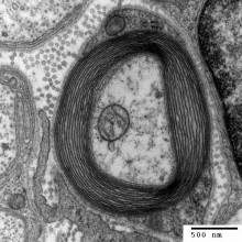
Previously, it was thought that the inappropriate immune attack was directed chiefly at a substance called myelin, fatty material that invests nerve fibres like the plastic insulation in an electrical cable. But now, and writing in the journal Immunity, Mainz-based researcher Volker Siffrin, from Johannes Gutenberg University, has discovered that a significant part of the immune assault is also directed at nerve cells themselves. 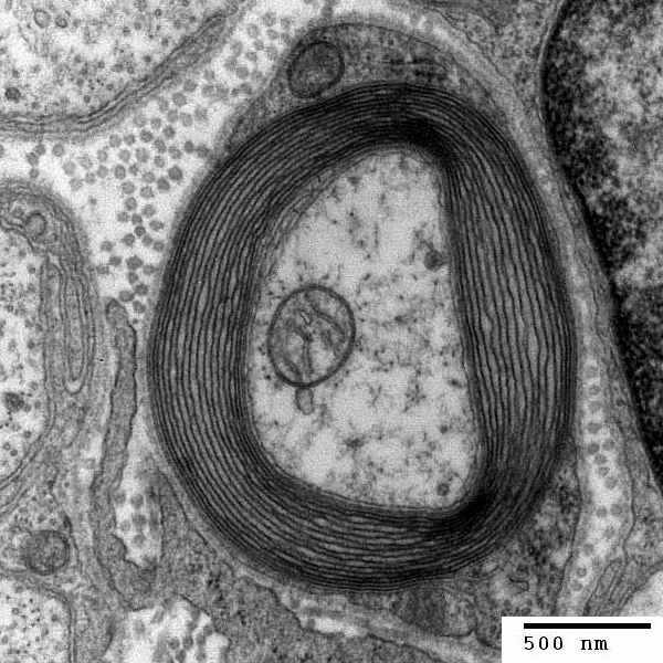
Together with colleagues, Siffrin studied mice with the rodent equivalent of MS that had also been genetically engineered to produce a red-coloured protein in their immune cells and a green-coloured protein in their neurones to enable the two cell types to be distinguished easily down the microscope. Using a technique called two-photon laser scanning microscopy (TPLSM), the German researchers carefully studied sites of inflammation in the brains of the MS-afflicted mice and saw a class of white blood cell, called Th17 cells, making direct contacts, called "immune synapses", with affected nerve cells both at the sites of inflammation and elsewhere along the course of the nerves in the brain. Contact with these Th17 cells, they found, caused calcium to flood into the nerves, triggering an injurious process called excitotoxicity, which can ultimately kill the cell.
So, far from being confined to the adjacent myelin, the underlying pathology of MS may well include simultaneous direct destruction of nerve cells early on in the disease course, accounting for some of the symptoms.
This suggests that targeting treatments at these immune-neuronal interactions could provide doctors with an additional weapon with which to combat the progressive destruction done by MS.

09:35 - Malaria: The Gorilla's Gift
Malaria: The Gorilla's Gift
with Professor Paul Sharp, Edinburgh University
 Historically, scientists thought that the most severe form of malaria, known as falciparum malaria, first spread into humans from chimps. But now, by looking for the parasitic DNA in faecal samples from thousands of wild apes, including chimpanzees, gorillas and bonobos, Edinburgh University's Paul Sharp and his colleagues have found that, in fact, it was gorillas that gave us malaria rather than chimps. He talked our own Smitha Mundasad through the work, due to be published in Nature this week.
Paul - Before we set out on this particular study, there was one species of Plasmodium known to infect chimpanzees and that's a species called Plasmodium reichenowe which is quite closely related to Plasmodium falciparum - the worst cause of malaria in humans. What we have subsequently found is that there are in fact six similar species of Plasmodium and we found three species of Plasmodium that only infect gorillas. When we compare those different species of ape Plasmodium to the human species, all the evidence suggests that the human Plasmodium has originated by a jump from gorillas to humans.
Smitha - What implications does that have now for our understanding of malaria?
Paul - Well, the initial aspect of this of course is just that it satisfies our curiosity about where humans acquired their Plasmodium parasite from in the first place. But ultimately, it can also prompt us, or others, to ask whether there are any differences between the strains that infect gorillas and that infect humans. For example, it looks like only one of those six species has successfully jumped into humans and it looks like it may have only done it on one occasion. These parasites are transmitted from ape to ape by mosquitoes and we would expect those mosquitoes also to be biting humans who live in close proximity to the apes. So we would expect there have been many opportunities for those Plasmodium parasites to infect humans but most of those occasions, they simply aren't successful. It looks like the gorilla strain that did make it into humans must've undergone some kind of adaptation - it would be really interesting to find out what that was.
Smitha - Could these findings lead in some way to find treatments in the end?
 Paul:: Any additional knowledge about what it is about these strains that allow them to infect humans or doesn't allow them to infect humans could lead to insights for therapies. Perhaps the most important implication of the work is that it says that there are these other species of parasites out there that might have the potential to infect humans in the future. And that would be particularly important if people were successful in curing humans of Plasmodium falciparum. There's a possibility that we're simply opening up the niche for one of these other ape parasites to jump into humans.
Smitha - Has this research thrown up any new questions?
Paul - There are all sorts of questions that arise out of this. We never find one of the chimpanzee parasites in faecal samples from gorillas and we never find one of the gorilla parasites in faecal samples from chimps. Presumably there's something about whether these parasites can successfully infect blood samples of other species, and it would certainly be intriguing to figure out - what is that species specificity? And of course, that then leads into the question of whether that specificity could change so that any of those parasites could infect humans in the future.
Smitha - Do you have plans for further research?
Paul - We do hope to look first of all at human samples to see whether any of these other ape species of Plasmodium have made it into humans. We also want to look further at the ape samples because Plasmodium falciparum, although it is the most common form of Plasmodium causing malaria in humans, is only one of four or five different species of Plasmodium that infects humans. We haven't really looked extensively at these ape samples to see whether the apes are infected by relatives of these other Plasmodium species that infect humans.

14:01 - New study points the finger at pain perception mechanism
New study points the finger at pain perception mechanism
In a breakthrough that might also help to explain why amputees suffer phantom limb pain, scientists have hit the nail on the head in explaining why we clasp our two hands together after we injure one of them.
UCL scientist Patrick Haggard and his colleagues, writing in Current Biology, used a trick called the thermal grill illusion (TGI) to fool participants into experiencing a painful sensation in one of their fingers. This clever trick involves placing the longest finger into a cup of cool water (at 14 C) and the two adjacent fingers into warm water (at 43 C). Paradoxically, subjects report that the middle finger feels painfully hot. 
This is probably because the warm sensations from the index and ring fingers partially shut off the cold sensations coming from the central finger. The reduction in the cold sensations then in turn makes the brain think that a different population of nerves, which signal painful heat, have become more active, so a burning sensation is experienced, despite the subject remaining totally uninjured.
Intriguingly, however, if the illusion is triggered in both hands simultaneously, and then the equivalent three fingers on both hands are brought into contact, finger pad to finger pad, the pain perception abruptly drops by over 60%. But this only works if both hands are subjected to the illusion at the same time, and does not work unless all three involved fingers are brought into contact, or if one of the hands is substituted by another individual. The researchers suspect that a specialised population of sensory brain cells, which integrate information from both sides of the body to produce a coherent body map, are responsible. "Correlated multisensory information is a major source of the sense of one's own body as a coherent ''self''. Our results show that coherence between hands, as well as across modalities, may contribute to pain modulation. The present study suggests that multisensory, multieffector information may rapidly boost coherence of cortical activity and thus reduce (thermal) pain," say the scientists.
Apart from its functional significance, this discovery could also have practical applications, such as in the treatment of phantom limb syndrome, a frequent complaint of amputees who experience painful sensations arising from their missing body part. The pain tends to reduce with time, perhaps as the amputee's brain re-creates a new coherent map. However, developing techniques to manipulate the map and achieve this new coherence sooner could help to reduce the duration of discomfort experienced by sufferers.

18:31 - Planet Earth Online - Physical Attractions in the Oceans
Planet Earth Online - Physical Attractions in the Oceans
with Helen Snaith, National Oceanography Centre, Southampton
A satellite designed to measure the Earth's gravitational field with unprecedented accuracy may sound like something out of a James Bond film, but it is in fact a reality. The European GOCE spacecraft or "Gravity field and steady state Ocean Circulation Explorer" has been doing just that and has recently sent back its first results. Richard Hollingham finds out more...
Richard - The launch of the dart-shaped GOCE satellite from the Russian Plesetsk Cosmodrome was in March 2009.
Helen - It is definitely the Formula 1 of satellites, if you like. It's designed to slip through the atmosphere as easily as possible.
Richard - Eighteen months later, in her office at the National Oceanography Centre in Southampton, Helen Snaith is studying the first data from this sleek satellite.
Helen - Its primary mission is to measure the Earth's gravity field at very small spatial scales. It's to see the very, very small changes in the gravity as you go around the Earth, and ultimately, to be able to use that information to get to ocean circulation.
 Richard - We'll talk about that in a second, but let's look at the first results. You've got some of these early results through, up on your screen here. It's an image of the Earth, but it's a lumpy, bumpy, multi-coloured, almost like a rock-like Earth.
Helen - This is an image that's been generated to show just how lumpy the Earth actually is. If you looked at the Earth, it's not completely smooth. We all know it's not completely smooth. We have mountains. We have oceans. We have valleys. But they have an effect on the gravity field as well. So the gravity field isn't the same all the way around the Earth. When you do maths, let's say in your school, you get taught that acceleration due to gravity is 9.8 metres per second squared. Unfortunately, that's not strictly true. There are very small changes as you go around the Earth. If you go over the Himalayas, there's more mass, there's more mountains, there's more gravity. So things are pulled towards them. If you go over deep trenches in the ocean, there's less mass, there's less gravity, and things aren't pulled towards them as much. It's these small changes as you go around the Earth that we're trying to measure with GOCE and which you can see exaggerated an awful lot on this image.
Richard - Okay, so you have a map of the Earth's gravity field which is what the satellite is producing. You want to use that to work out ocean currents. How do you make that leap?
Helen - Because the gravity field isn't the same everywhere, the water in the oceans isn't being pulled down to the same level everywhere. So, if the water was completely still and there were no ocean currents, it would be a bumpy surface. But if you put a ball down on that surface, it wouldn't roll anywhere. It would stay where it is, which is a slightly strange concept to be able to get your head around, but this is where we're coming from. What we're trying to calculate now is the difference between that nice smooth steady state - that if there were no currents, this is the shape of the sea surface - and what we actually measure - the difference between those two is what's being caused by the ocean currents.
Richard - So the only way you can work out really the impact of the ocean current or the height of the water as a result of these ocean currents is by removing the gravity from that.
Helen - Exactly. That's precisely what we want to do. We can use an altimeter - satellite altimeter - to measure the precise height of the sea surface at any given time when the altimeter flies over. What we don't know is how much of that height is being caused by the gravity field or that change in height. With GOCE, we can start to get a handle on that. That's what we're really interested in.
Richard - Okay, you're interested in it. Why is it important to learn or work out where the ocean currents are and what effect they're having?
Helen - The ocean currents are a really important part of the heat transport system of the ocean. What we're trying to do is to monitor those currents and see how consistent they are, how strong they are, whether there's changes in those currents. To be able to do that, we need to be able to monitor them globally. Satellites give us one of the few options we have of being able to look at the currents everywhere in a consistent way.
23:16 - Mind-reading MRIs
Mind-reading MRIs
with Professor Jack Gallant, Helen Wills Neuroscience Institute, UC Bekeley
Chris - Thank you for joining us on the Naked Scientists. Can you tell us first of all, what actually is fMRI? How does it work?
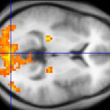
Jack - Well fMRI measures brain activity, but it does it rather indirectly. It doesn't measure the activity of neurons in your brain. Instead, it measures changes in blood flow in your brain. So, when your neurons fire, they need to use energy and they get their energy by burning glucose with oxygen, and they extract glucose and oxygen from the bloodstream continuously as you think. And the areas of your brain that are more active, meaning more neurons are firing, tend to extract more oxygen and glucose from the bloodstream and we can measure this [blood flow] using MRI.
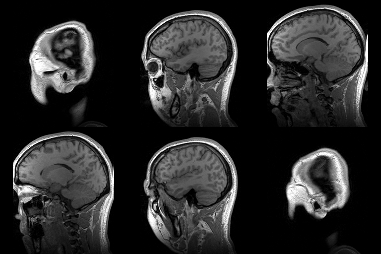
Chris - In other words, it's a correlate of what the brain is doing so when we present a stimulus at the brain, and we look at which bits of the brain are using more oxygen through the signal in the fMRI scanner, that tells you that bit of the brain must have something to do with the stimulus that we're presenting and how it's being decoded.
Jack - Right. It's a rather indirect measure of something that's correlated with neural activity and it's the best measure we have right now, the best non-invasive measure of brain activity, but the problem is that it has fairly low spatial and temporal resolution, relative to the neurons themselves. So you're losing a lot of information or you can't recover a lot of the information that's actually going on in the brain. Still, it's the best tool we have at the current time.
Chris - So actually talk us through what it is that you're doing and how you're analysing the signal that comes back from the brain? What sort of resolution can you get?
Jack - Well the typical functional MRI experiment that people do today will recover information from a small area of the brain - a small resolution of the brain in about 3 x 3 x 3 millimetres. And so, your brain is essentially divided into a large number of cubes - tens of thousands of these little 3 x 3 x 3 millimetre cubes called 'voxels' and for each individual voxel, we can build a model that describes how your brain encodes information. So for example, if we put you in a scanner and we show you pictures or movies, we can construct these models called 'encoding models' and each small little voxel in the brain will have its own unique encoding model that describes how the movies or the still images are translated into changes in blood flow in that small region of the brain.
Chris - And what does that actually tell you about the underlying structure of the brain in that region, the fact that you've got these little cubes and they're changing their activity? What can you infer about brain activity from the patterns of activity that you see in the scanner?
Jack - Well, the encoding models essentially tell you how the external world is translated into changes in blood flow. So if you show someone thousands and thousands of still images or say, a few hours of movies, you can actually build a model that's quite general and it describes how any possible visual stimulus that you could show that person gets translated into blood flow. Now once you have that model, you can then demonstrate that it's working by using it to identify on each individual second which movie or which picture they saw. Now once you verified that the model is accurately identifying the image or movie they saw, you can actually show the subject a completely new movie or image that they never saw before and you can actually reconstruct that movie or image from the blood flow that you've measured.
Chris - Does this give us clue though how the brain is actually wired up, so when you look at how the brain responds to a picture you show, does this inform the way in which it's decoded or deconstructed cognitively to then present to consciousness, what we're seeing when we experience that stimulus?
Jack - Well absolutely. In fact, that's the entire point. The brain processes visual information with a large number of different brain areas. There's probably something between say, 50 and 75 different brain areas that are involved in visual function, and the goal of our lab is actually build computational models that describe how all these different brain areas work. So, when you undertake to build these encoding models, what you're really trying to do is construct a quantitative theory of the way the brain processes visual information. And decoding is simply a way of verifying that your theory is actually correct. If you have a correct encoding model that describes how information is translated into patterns of brain activity, then you should be able to decode it accurately and reconstruct the stimulus that this person saw. But that's sort of a side effect and it's actually just a mathematical trick. The main goal is to build an accurate encoding model and once you have that, decoding sort of comes along for free.
Chris - And if I compare how the brain of one person responds to a picture postcard and then I present the same picture postcard to a second subject, do you get broadly the same activity in the brains of the two individuals? In other words, could you build a system that will pretty well work out what they're seeing or is it so end-user specific that you'd have to train the system to look at each individual in order to do it with any particular level of accuracy?
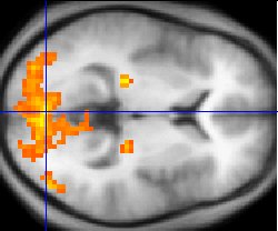
Jack - Well the answer is somewhere in the middle there. Individual brains vary in huge amounts, just like individuals have different heights and different weight. The size and shape of individual brains varies a lot. So if you build a sort of generic model that you can apply to any brain, you will be able to decode some coarse information, but the model will never work particularly well. If you want to recover a lot of information from someone's brain, you're going to have to build a unique model for each individual person's brain.
Chris - So tell us about the experiments that you've actually done to show that this can be done. It can be done successfully.
Jack - Well our published experiments have involved static images and the procedure is pretty simple. We put somebody in the magnet and we show them 2 to 5 hours of images. These are just flashed every few seconds and they passively view them and after we get a large enough data set, then we can construct these encoding models on a computer and then we simply put them back in the magnet and we show them new images they've never seen before, and we use the computers to reconstruct the images that they actually saw. In our more recent work that has not yet been published and is still in peer review, but we've shown that you can do this with movies as well and that was kind of an interesting challenge because the blood flow signals measured by fMRI are very, very slow. You're only getting a snapshot of the brain once every 1 to 2 seconds. And at that rate, it's actually quite a challenging problem to try to reconstruct the motion of natural visual stimuli.
Chris - So how are you getting around that? Are you going for things like, if there's a very, very apparent thing in the movie which triggers a certain response in that person, say the person sees a post box or a cat and their brain is going to respond strongly to the post box or the cat, is it that that you're picking up rather than the fact that you've got a moving image going across and lots of stimuli being presented sequentially?
Jack - It's actually both. So, your brain has probably something on the order of 50 to 75 distinct visual areas. No one's really sure exactly how many visual areas there are. What you ideally want to do is build an accurate encoding model for each individual visual area and that encoding model will describe the way information is processed in that specific visual area and will describe the features in the natural images or natural movies that are essentially represented explicitly in that area. And once you have those individual models, then when you do decoding, you aggregate information from all of these different visual areas. So visual areas that are sensitive to motion, but they don't care what the moving object is, then you'll reconstruct the absolute motion from those areas. Other visual areas that may be more involved in semantic information like they may respond to people talking or to vehicles moving, they'll reconstruct the semantic category from those areas and then you aggregate all that data together to produce a reconstruction.
Chris - And if you can get this working at an even better resolution you do at the moment, what are the big questions that you now want to go on and answer with this tool?
Jack - Well again, the main goal here is to build encoding models and what we want to do is to be able to perfectly predict all of the brain activity in as much of the brain as we can. So, we haven't gone particularly far in that part yet. We are still in the process of building more and more accurate encoding models especially for that more mysterious visual areas and that work will be going on for quite some time. But all of the mathematical algorithms we use for constructing the encoding models and for doing decoding can be applied pretty much anywhere in the brain. So for example, you could imagine building encoding models for auditory areas, for recovering music and speech processing. You could imagine building these models for the frontal areas of the brain that are involved in abstract thought and that work will be going on for a long time because there are few computational theories that neuroscientists have right now that describe accurately how the higher order sort of more mysterious parts of the brain actually work. Right now, we're working at the visual system because it's relatively easy, relative to all of the other parts of the brain. We all know what visual system does, we have some rough ideas of how it's laid out, and we can control the input very carefully. If we try to build a model of the front part of the brain where you think about your future and you plan, and all those sorts of things, it's both very difficult to model those areas and it's very difficult to control the input to those areas. So, progress in building encoding and decoding models for more abstract parts of the brain is going to proceed much, much more slowly.
33:28 - Hallucinations and delusions - what's going on in the brain?
Hallucinations and delusions - what's going on in the brain?
with Professor Paul Fletcher, Department of Psychiatry, Cambridge University
Kat - So if you could tell us a little bit about what fMRI can actually tell us about what's going on in the brain and what it can't actually tell us yet? I think some people think it's maybe a magic mind reading machine. Where are we at the moment with this technology?
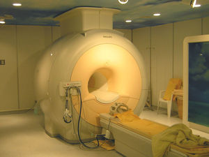
Paul - Well, as we were just hearing from Dr. Gallant, fMRI is a way of looking at which different bits of the brain respond when people are asked to do things or when they're having particular experiences. From my perspective, it's very interesting because we can start to look at the areas of the brain that become active when somebody's experiencing quite unpleasant symptoms that co-occur with mental illnesses. So for example, a hallucination where somebody is perceiving something that isn't actually there; for example hearing a voice when there's nobody around or seeing something that isn't there. Or delusions where they believe very strange things, often very unpleasant things such as their neighbour is trying to kill them or something. And fMRI offers us a way of putting them into the scanner and looking at how their brain is behaving in this sort of setting.
Kat - So, how are you actually doing this in practical terms? What's your research involving?
Paul - Well the first thing is, I'm not actually terribly interested in trying to map the hallucination to the brain. I think that would be a lovely thing to do, but the problem is, if you lie somebody in the scanner, and they're having hallucination, you can get them to indicate when the hallucination occurs, and you can watch what changes in the brain are occurring, but you don't actually know whether those changes are the course of the hallucination which would be very interesting, or whether they're actually a consequence. For example, if I'm hearing a voice, my brain will respond to that voice even though it's not causing that voice. Or there may be a compensation; so, a hallucination would be very unpleasant and that the changes we see in the brain might actually be a result of somebody trying to suppress the unpleasant thoughts and connotations. So I'm much more interested in trying to use functional imaging to try and test particular cognitive or psychological models of how the hallucination occurs, what computations in the brain may be disturbed as a prelude to hallucinations or delusions.
Kat - So you're trying to sort of strip out some of these variables. How are you actually doing that?
Paul - Well, we're taking as our starting point an acknowledgement that actually, pretty much all of us live in a fantasy world where we are hallucinating quite a lot of the time. We're creating our own reality. The reason being that if we were to try and sample all of our neural inputs at once, all of the sensations that are impinging on our brain, we would be totally paralysed by information. And a consequence of that is we start to take shortcuts and we start to process the world, not so much as a result of what is there, but of what we expect to be there. And I think we can think of our brains as being a very delicate balance between what we're predicting to see, or hear, or feel, and what we're actually experiencing through our neural apparatus. And my belief is that we can start to understand some of the symptoms of mental illness by looking at this balance and by acknowledging that there's a very important signal in the brain that almost pervades all neural firing which is the mismatch between what we expect and what we've got, the so-called 'prediction error.' And so, what we can do is look at how prediction error firing in the brain occurs in people with and without hallucinations and see if that is disordered in some way. For example, do they appear to show surprise or prediction error by things that should actually be highly predictable or contrarily, do they show an absence of prediction error when there should be something surprising occurring. So we can use imaging to test this and in so doing, try and understand the basis of these experiences.
Kat - So I understand that you're using a drug called ketamine to try and make people hallucinate. My friend have this when he broke his leg. Doctors gave it to him and he said that the doctors were fine putting his leg back together, but they were all talking backwards. What can that tell us about hallucinations in the scanner?
Paul - Well, the big advantage of ketamine is that you can control it. You can control the dose that somebody gets. You can infuse it, you can keep them at a particular level for a certain amount of time, and you can watch how their brain responds. You're not reliant on the chance and randomness of normal hallucinatory experiences. So we use ketamine as a way of producing very temporarily these experiences and seeing what it does to the brain as a way of trying to understand how the hallucinations or delusions might arise in the context of mental illness.
Kat - And so, what do we think about what is going on in mental illness when people are having these hallucinations and delusions? What's actually going on in their brain? What have you found out so far?
Paul - Well what we seem to be finding is that people with these experiences are actually getting false signals in their brain that the world around them has become surprising, and is sort of baffling their expectations. They can have very strong expectations about what they're about to see or hear, and even if what they do see or hear actually fulfils those expectations, nevertheless, their brain is telling them that it is wrong. And so consequently, they have to keep changing their predictions, rebuilding their models of the world, trying to understand the world in new, and evermore often bizarre ways. And I think there's good evidence emerging across our work and a series of other studies elsewhere that this may be a fundamental deficit in certain mental illnesses.
Kat - And so, really briefly to touch on this, we sometimes hear about some great creative artists, very creative people who are also judged to be mentally unstable or mentally unwell, do you think maybe that some people who are extremely creative are slightly unhinged in this way that their hallucinations are maybe coming to the surface more?

Paul - I think that's a very interesting point and certainly, people have played with that idea. My own feeling is that up to a point, the sorts of processes that might be deranged in hallucinations and delusions, up to a point, actually, having a slight derangement in those could be advantageous because it might lead you to look at the world in very new, and salient, and original ways that might actually give you insights that perhaps you wouldn't normally get by just predicting the world and it always fulfilling your predictions. So I think there may be a link there, but I have yet to actually draw it and so, I wouldn't want to speculate too much.
Kat - Absolutely fascinating then, a line between genius and madness. That was Cambridge University's Professor Paul Fletcher.

55:00 - Are Apple Cores Poisonous?
Are Apple Cores Poisonous?
Diana O'Carroll put this question to John Fry, a consultant in food science...
John - Well, it could be but only under rather extreme circumstances. Apple seeds contain a substance called amygdalin that can release cyanide under the right circumstances such as contact with digestive enzymes. The cyanide is linked to sugars in the form of a cyanogenic glycoside and these cyanide-releasing compounds are remarkably common in nature. They occur in more than 2,000 plant species, some of them important foods like cassava. They also crop up in stone fruits like plums, peaches, apricots, and famously, bitter almonds. It's often said that cyanide smells of bitter almonds, but actually, it's the other way around; bitter almonds smell of cyanide.
You need about 1 milligram of cyanide per kilo of body weight to kill a human being. Apple seeds contain about 700 milligrams of cyanide per kilo, so about 100 grams of apple seeds should be enough to dispatch a 70-kg adult human, but that's an awful lot of apple cores even if you don't eat the rest of the apple first. In addition, the seeds would have to be pretty finely crushed to let the enzymes get to the amygdalin at all. All in all, you're safe eating the occasional apple core. I've done it for years. Just don't try eating a bowl of freshly crushed apple pips.
Diana - If a seed weighs 0.7 grams, then you'd need to munch your way through 143 seeds. Apples can contain anywhere between 2 and 20 pips, but a typical supermarket apple will contain about 8. So you'd have to eat about 18 apple cores in one sitting!
- Previous Nerves in Cancer, MS and Pain
- Next Making Steam Inside Stars


Comments
Add a comment