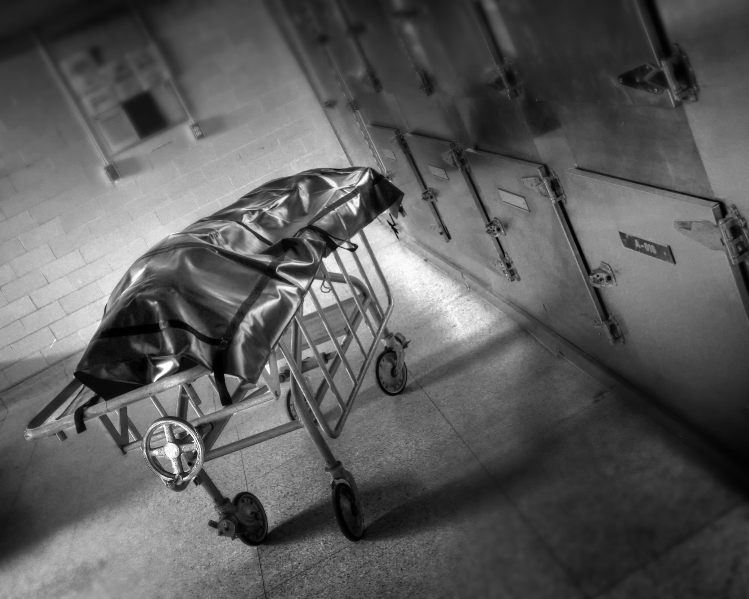
Autopsies: determining cause of death
Interview with
Post mortems are carried out by forensic pathologists; they look first at the external surfaces of the body to check for signs of trauma, then they examine the internal organs to piece together what might have caused someone's death. Jocelyn Pryce from Anglia Ruskin University demonstrates to Chris Smith, using pig organs, what a pathologist would be looking for...
Chris - So, what would actually happen when our gentleman down here who's our victim turns up in the post mortem lab? What's the process? What do you do?
Jocelyn - The most important thing would be that we could identify the body. So, we could ensure that it's the same one from the crime scene that's actually arrived in our mortuary. This is important because we need to be able to trace it. We need to know who's had access to him and who could obviously have planted evidence along the way onto the body itself.
Chris - So, you get him on the slab. What do you look for? I know I said that you look for external signs of things, but what sorts of things can you pick up by just looking at someone from the surface?
Jocelyn - We can get a lot of information by doing a visual examination of the body. The clothing would then be taken and exhibited separately. And then we would start to actually look at the body itself. We would look for things like bruising, defence wounds on the forearms, defence wounds seen on the hand, on the palms of the hands, if somebody's been attacked by a knife, bruising around the head. So, all those sorts of things would be important for us to take account of during our examination.
Chris - Can you also get some idea as to how long someone's been dead because when you watch telly programmes and the detective always says, "When was the time of death?" And this person gives a prediction almost to the nanosecond on telly, don't they? I mean, it's obviously not as accurate as that, is it?
Jocelyn - There are guides for us to be able to tell how long a person has been dead. We can look at the amount of decay if they've been dead quite a long time. We can also look at things like rigor mortis which sets in over certain periods and then wears off again. So, that enables us to pinpoint time of death.
Chris - So, the person goes a bit stiff and then they soften up. So, if they're still stiff, you know that they've died sooner rather than later.
Jocelyn - Yes, they actually go very stiff and that will wear off over time. So, we've got a fair idea then of the timeframe that we're talking about.
Chris - What about simple things like just taking the temperature because obviously, we're a big bag of water, aren't we? So, it does take quite a while for a human to cool down.
Jocelyn - Yes. It very much depends on the situation that they're found in and the temperature of their surrounding areas. So for example, we would see advanced decomposition in somebody in a flat where the heating have been left on quite high. They wouldn't have necessarily been dead for as long as we would have expected. The rate of decomposition would be much, much faster.
Chris - What about telling if our victim had been moved? Can you tell that by looking at them externally?
Jocelyn - Yes. Again, we can pick up a lot of information about whether they've been moved, what they've been leaning on, the way that the blood pools in certain areas, where there's been a lot of pressure. So for example, if somebody is laying on a mat with some indentation in it or something like that. Quite often, you can actually see that where the blood is pooled onto the body. You can actually pick up the patterns. So, it's very important to have a really good visual examination of the body.
Chris - What about when you get inside?
Jocelyn - Okay, so after we've made the visual examination of the outside of the body, we would then consider the internal organs. As an example, what we've got here in this tray, we have - it's actually a pig pluck - we're using a pig for this.
Chris - I'm relieved because there is sitting in front of us, on this very large black tray, a whole load of what I would call the offal group of organs. I was wondering where you've got them from because knowing as I do a human pathology then they look pretty similar.
Jocelyn - Yes. This is actually what we call a pig pluck. So, it consists of the lungs, the trachea, the oesophagus, the liver, kidney, and heart. And can buy them from a butcher, they will come all in one piece. You can buy them and we've dissected this one out slightly so that we can have a look at their separate organs.
Chris - They taste great in haggis. So, if you were actually to be faced with the remains of the internal organs of our victim, how would you then approach looking at these organs? What are you looking for?
Jocelyn - Well, in this particular case, we obviously don't know yet how he's died, the cause of death. So, we would be looking at maybe the pathology because there's no obvious signs of death. So, we'd be looking at pathology, we'd examine the heart, see if there's any signs of a heart attack. We'd look at the lungs to see if there was congestion. Yeah, we'd have a good look around all the internal organs and see how normal they look. Obviously, these all look fairly normal, but there would be signs if there was a heart attack. There would be signs in the heart muscle that the blood had been deprived in a dead area of muscles so it would be obvious to us.
Chris - If our man had been poisoned, how might that show because obviously, I can understand, if you've had a heart attack, you might see a blood clot in a coronary vessel for example and as you say, bits of dead muscle. But how would say, the organs be affected by poisoning?
Jocelyn - It very much depends how somebody has been poisoned. How it was ingested, if they ingested it, you could find evidence of irritation in the trachea, you could find evidence of irritation in the intestines, lungs, liver damage if it was over a long time. so, it very much depends on how the poison was actually taken into the body.
Chris - And therefore, which organ gets hit the hardest.
Jocelyn - Yes.
Chris - You've got some little pieces cut-off here. Is that because when you did the sort of gross examination of the organs, you then take some smaller samples for further examination?
Jocelyn - Yes. So, when we have a look, what we would do is remove the sample, remove the organs here and we could bread slice them and actually have a look at the cut surfaces all the way through to identify any areas of damage.
Chris - We're looking at the lung here and you cut the lung into sort of 1 centimetre wide lumps.
Jocelyn - Yes. That's so we can have a look all the way through it and we would do exactly the same with the heart. We'd open up the heart and have a look at the walls, the ventricles, the atria, and have a look all the way around to see what sort of damage there was. So, if for example somebody had been stabbed then we might see damage in the lungs or in the heart, the evidence of the stab wound.
Chris - How do you actually record your findings? Do you have to take photographs so that when Alan needs his evidence, you're able to show him the physical pictures of what this person look like inside to account for their death?
Jocelyn - Yes. We would take photographs of anything that we found and also, there would be a report constructed of everything that we found, so yeah.
Chris - So, let's turn one of our audience into an amateur pathologist. What's your name?
Nicole - Nicole.
Chris - Nicole, you've suited and booted. That's lovely and you've got purple gloves on. That's terrific! So, what would you like to find out about first?
Nicole - Let's have a go at the heart. It's quite dense. It's quite heavy.
Chris - How much do you think that weighs?
Nicole - A couple of pounds?
Jocelyn - She's probably about right. Yes, it's a small but very, very dense, very heavy organ, yeah.
Nicole - Compact muscle and you can see the different compartments as well quite nicely, I guess.
Jocelyn - So, we've got the atria here at the top and then we've got the ventricle lower down. Obviously, it's very dense because it's designed to pump the blood around the body, so it has to be very strong to be able to do that.
Nicole - So, this would be the same size as a human heart approximately?
Jocelyn - Slightly bigger than that, but yeah.
Chris - What else can you see there, Nicole?
Nicole - So, these are the lungs.
Chris - Is that what you expected a lung to look like?
Nicole - Well obviously, it's chopped up so I would hope my lungs wouldn't look chopped up. But it's very light. It's a lot lighter than I thought they would look, a lot less dense muscle. Yeah, I guess I would imagine the lungs to look a bit like this as a whole.
Jocelyn - It's a lot lighter because it's full of air. So essentially, it's just lots and lots of very tiny balloons with very fine membranes because it's obviously involved in gas's exchange. So, they are very light. They're much bigger than the heart but they're much, much lighter because they're full of air.
Nicole - Yeah, very squidgy.
Chris - Thank you very much, Nicole. Give Nicole a round of applause please. What about if you do all these and you think, "Well, to all intents and purposes, these organs look pretty normal and I can't see any signs of trauma." What do you do next?
Jocelyn - In the case of our young man down here, what we would do is we would submit blood samples to the laboratory and also the stomach contents to the laboratory as well so it would go off and be dealt with separately away from the mortuary.
- Previous What's your poison?
- Next At the scene of a crime


Comments
Add a comment