Intelligent knife sniffs out cancers
Interview with
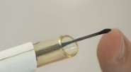
 A new intelligent surgical device that can chemically analyse the tissue it's cutting through to distinguish between healthy and cancerous cells have been unveiled by scientists at Imperial College, London. Chris Smith spoke to co-inventor, Zoltan Takats to find out how the "iKnife" works...
Zoltan - It is actually a hybrid instrument. It is a combination of a surgical dissection devices which is actually a quite old surgical device called electrosurgery. It's been used since 1925 I believe. So, it's a combination of electrosurgery with an analytical device called mass spectrometry. We are taking the surgical dissection tool and using the by-product of the surgical dissection tool to identify what kind of tissue the surgical dissection tool is cutting through.
Chris - When one does electrosurgery, effectively, you're using an electric current. It generates little spark at the tissue interface and that vaporises the tissue. Having done this, I can testify to the smell that comes off. There is a lot of smoke. You're presumably drawing off some of that smoke and then feeding it into your mass spectrometer to sniff the smoke and see what's in it.
Zoltan - Exactly. Electrosurgical device produces this smoke. This aerosol and we realise that this smoke doesn't only contain the tissue debris and the things like that, but it also contains ionised molecules. So molecules that are actually charged and without doing too much to this aerosol, we can introduce it into a mass spectrometer and analyse the charged molecules, and get a chemical fingerprint of the tissue which is being dissected.
Chris - In other words, when you blast apart the cells with the electrical cutting tool, effectively, you're also releasing as a vapour, all of the chemicals in those cells, and I suppose the premise here is, if a cancer cell has a different biochemical milieu going on inside it compared with a non-cancer cell, can you tell them apart?
Zoltan - So, the cancer cells have a markedly different chemical signature and that's what we use to identify them.
Chris - What sort of resolution and how would this be useful for a surgeon?
Zoltan - Resolution wise, it always depends on the hand of the surgeons. If you look at the instrument or resolution that we can actually look at micrograms of tissue in using this method Of course, surgeons cannot really work with micrometre precision. I would say, you're using a handheld device, maximum resolution is somewhere around a millimetre but it clearly depends on who is holding this device.
Chris - So, would one apply this in a sense that a surgeon has operated on a cancer, the surgeon thinks that he or she has got all of the tumour out, but by running the device around the margin of the tumour, and sniffing that smoke that's coming off, this will tell if there's still residual cancer tissue there or if they're into healthy tissue?
Zoltan - One thing is when a surgeon is cutting around the tumour and he or she assumes that all the dissection line is healthy tissue, the device would cut through tumour or cancer environment, so something which is not cancer but really close to the tumour, then the device can give warning signal, the identification result on the screen. We have actually a very simple colour coding with cancer in red and tell the surgeon that maybe you want to remove a little bit more tissue here because you are getting too close to the tumour. But if it's a wrong type of application, another type of application that would be the identification of unknowns, fairly often it happens that there are such tissues, unidentified tissue features around the tumour and that it's really hard to tell just by the naked eye whether these are proxima metastases of the tumour or these are just something else. In these cases, it is very important thing to do is decide because if it is a metastatic disease then there is not too much hope for a curative intervention. If it's not metastasised yet then there's a good chance to cure the disease. At that point, when the patient is opened up, then surgeons can see what's around and has to make a decision which way to go. But in our case, one can just zap a little bit of the suspicious tissue, a hardly visible amount and get an answer immediately.
Chris - Now, when one does a resection, the pieces of whatever has been taken away go off to the histopathology laboratory and a pathologist looks down a microscope to see if they can see where the unhealthy tissue stops and a healthy tissue starts to gauge this so-called resection margin. Could the histopathologist, instead of relying on his or her eye on a microscope, could they use your tool to ask, does this thing have clear margins? Where does the tissue become unhealthy again?
Zoltan - Actually, you've pointed out the very important feature here. This intelligent surgical device is a part of a much bigger story and this much bigger story is about paradigm change in histopathology. What we are proposing here and not only me, but also a number of other groups all around the planet is to change the basic definitions of tissues from a morphological basis to a chemical basis. In that sense, histopathologists can use not surgical device, but can use similar tools with higher space or resolution which we generally call imaging mass spectrometry for the
identification of tissues in surgical sections. But that's again in the area which we've been heavily working on, but that's a little bit different from the current study.
- Previous Ketamine and schizophrenia
- Next Inside eLife, July 2013


Comments
Add a comment