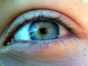
Seeing Red - Blood vessels in the eye
Interview with
There are at least two places in the body where blood vessels are actively  discouraged from growing: one is the cornea, the other is the retina. And with good reason, because if blood vessels do invade these sites, they can cause blindness. But how is this achieved? It turns out that both sites produce a soluble molecule that soaks up - and therefore blocks - the signal that would normally trigger blood vessels to form. Chris Smith spoke to Bala Ambati from the University of Utah...
Bala - When you look at the eye, it has several important yet, almost contradictory missions. The cornea in the front of the eye has to stay clear, it has to stay strong, it has to focus and it has to do all that without having any blood vessels that can give it nutrition or oxygen. Now in the retina, at the very back of the eye is what's called the photoreceptor layer and the photoreceptors are the rods and cones that convert light into sight. Those rods and cones use a lot of oxygen. They're actually the most metabolically active cells in the body. And so, they are situated right next to the choroid - the blood vessel tissue network that has the highest blood flow of any vascular network in the body. And so, you have these photoreceptors that demand high oxygen, situated right next to this incredibly rich blood vessel network. Yet, that layer of the retina should not have blood vessels because as soon as blood vessels come in, that can distort the vision. And so, you have these contradictory imperatives - clarity versus oxygen demand.
Chris - How did you approach that and begin to ask, what keeps the blood vessels out of the retina, despite its very high oxygen demand. and equally the cornea?
Bala - I started looking at the cornea and realised that the cornea expresses several different VEGF receptors and VEGF is a vascular endothelial growth factor, but interestingly enough, it expressed one particular form of VEGF receptor called sFlt-1, soluble VEGF receptor-1. We looked at multiple mammals. One of the very few mammals that in nature, does not have a clear cornea is the Florida manatee and it actually has a vascularised cornea. A couple of mouse strains that are genetically mutated also have vascularised cornea. And so, we found that in these mutant mice and in the manatee, this sFlt-1 was missing. And we also found that restoring sFlt-1 to mice that were lacking in the molecule helped restore corneal clarity and the lack of blood vessels.
Chris - Is the model of a hypothesis then that the cornea in this instance is secreting this molecule which has the effect of soaking up a factor which would otherwise trigger the growth of blood vessels? It's locking it away, so that the blood vessels do not grow.
Bala - Precisely.
Chris - And so, then I guess, you must've thought, well, if that's what's going on in the front of the eye, could the same trick be playing out in the back of the eye, in the retina? So, is there a soluble form of this - for want of a better phrase - blotting paper for VEGF, the factor that makes blood vessels in the back of the eye, preventing it from acting there in the retina too?
Bala - Absolutely. So, the soluble VEGF receptor is a decoy - or as you put it, blotting paper - so it serves as a sink for VEGF. And so, our very first preliminary experiment was just to see, well is this molecule expressed in the retina? And interestingly enough, it was expressed in the retinal pigment epithelium which is the layer that is underneath the rods and cones, and just above the choroid. And so, that thin red line if you will, of RPE tissue is what keeps those choroidal blood vessels out of the retina and the RPE does indeed express sFlt at high levels.
Chris - So, I suppose it could be regarded as the million dollar question - if you look in people who've got disease in their retina, diseases characterised by new blood vessels growing in - things like wet macular degeneration, do they lose the secretion of this VEGF blotting paper, the sFlt signal which allows those blood vessels to invade in the way they do pathologically?
Bala - Exactly. I'd say, that's not just a one million dollar question, but it affects 10 million Americans and 2 million Britons. And so, we've proceeded to examine what happens in patients with disease then we did a number of experiments in mice, and we found that knocking down this molecule sFlt, allows these blood vessels, the choroidal blood vessels to invade the retina.
Chris - Do you think that if we were to go in and therefore turn that signal on more or enhance it in people with disease, we could arrest the disease process and do it in a safe way?
Bala - Correct. There's a sister paper that came out in ACS Nano, just a couple of months ago where we present the data on that exact same question. We found that in both a mouse model of macular degeneration and a monkey model of macular degeneration, that if we delivered nano particles loaded with a subunit of Flt-1, inject it intravenously, not into the eye but just their systemic vein, it could cause regression of these choroid blood vessels growing into the retina in both mice and monkeys.
- Previous Can science combat stigma?
- Next Building with Mushrooms


Comments
Add a comment