In this episode of the eLife podcast we discuss doing protein crystallography with electrons, the discovery of a receptor for carbon dioxide, new insights into arthritis, how the brain responds to a missing hand, and the best shape for whiskers.
In this episode
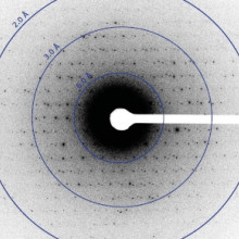
00:39 - Gently does it for crystallography
Gently does it for crystallography
with Tamir Gonen, Janelia Farm Research Campus, VA
Very cold, weak electron beams have been used to collect large numbers of diffraction patterns from protein microcrystals...
Tamir - We study proteins that live in the surrounding membrane of a cell. They form gateways for sugars and other molecules like ions and water to go into a cell or out of it, and we arte trying to study how they're built and based on how they're built we're trying to understand how they work and what goes wrong in disease. And one of the major problems that we've had in the lab is that to form crystals from which you can deduce a structure is very difficult for such membrane-embedded proteins. And so, often what we got were crystals that were way too small for structural studies. And what we were trying to figure out is, is there another way of getting to the protein structure out of crystals that are really very small?
Chris - So, how were you trying to image them when you were making these crystals or, all be it, small ones? How were you then trying to get the structure?
Tamir - The most common way of determining a structure from a crystal is by a method called x-ray crystallography. Now, you can think of a crystal as a three-dimensional object that has repeat structural motifs in it and when you shoot an x-ray beam through it, those repeat structural motifs act like a diffraction grating and they split the x-rays and scatter them in a predictable way. And based on the scatter that you get, you can then ask what kind of a structure would give me such a pattern?
Chris - Is the downside that you need a lot of crystal matter there in order to get that reproducible pattern in order to deduce what it's made of?
Tamir - The downside is that the x-rays have a lot of destructive energy in them because it's such a strong beam. So you need large crystals for x-ray crystallography to be able to withstand this large amount of radiation that we're giving it.
Chris - So, how have you tried to get around that?
Tamir - Well, so what we tried to do is rather than using x-rays, is using electrons. And the thinking behind it was that electrons interact with matter much better than x-rays, and so maybe we could get away with using much smaller crystals.
Chris - Why has no one done that before because we've had electron microscopes for donkeys years, people have been imaging many, many things in enormous detail with electron microscopes? So, why had no one tried to do what you did?
Tamir - People have tried to do that before. Many of my colleagues have put crystals in an electron beam before. The issue is when people use electron microscopy for biological specimens, they use a procedure that's called, low-dose cryo-EM. That means that they try to limit the amount of electrons that hit the sample, because if too much electrons hit the sample, then just like with x-rays, the sample will get destroyed. The low-dose procedure limits the dose to about 20 electrons per square angstrom. Now, if you take a crystal and shoot it at that dose rate of 20 electrons per square angstrom, the crystal gets destroyed after a single shot. So, what we said is, well, if a single shot at 20 electrons per square angstrom would destroy the crystal, what would happen if instead of using 20 electrons per square angstrom, can we use 10? Would we still get meaningful data? And the answer was, yes. So then, we got greedy and we said, okay, well can we cut it down more? And we got it down to 0.01 electrons per square angstrom per second. And so, that's about 200 times less in dose than what my colleagues were using and yet we still got meaningful data up to what is called, atomic resolution. Once we knew that we can get so much data out of a single crystal, all we had to do is that while we're exposing it, we start tilting it. And it's that tilt that gives you the different views, if you like, the different patterns from all the different angles and because you have all these different angles in three dimensions, you can calculate back and get a structure.
Chris - And what sort of quality of image can you get out because, obviously, we can get some idea of the crystal's structure with an x-ray, a pattern? But with electrons, is there anything else that you can see beyond just working out what the crystal structure is?
Tamir - Well, in x-ray diffraction, what you end up with is, is an electron density map. In microED, what you end up with is a column potential map, and so that means that you could look at charge. So, if you have a process that is charge-coupled, for example a proton-coupled transporter, you could figure out which amino acid residues in a protein get proteinated or deproteinated. In other words, you can see where the charges are. And that would really begin to help you explain the mechanism by which a protein works, and that you can't do by x-ray crystallography.
05:57 - CO2 sensor: if blood's too acid, you die
CO2 sensor: if blood's too acid, you die
with Louise Meigh, Warwick University
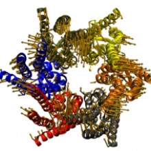
A nerve cell, connexin, can detect carbon dioxide, and could be involved in processes as diverse as breathing, 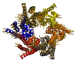 hearing and reproduction.
Louise - You need to monitor the amount of CO2 in your blood. CO2 reacts with water to make hydrogen ions, so it makes the blood more acidic and because enzymes are sensitive to that, if your blood gets too acidic, then you'll die.
Chris - So, how did scientists and doctors think that CO2 was registered prior to you coming along?
Louise - So the main theory is that it's by pH, so that's measuring the amount of these hydrogen ions. And there's lots of evidence for that, and it's definitely involved. But there's more evidence that CO2 is monitored directly as well, which is what we're looking into.
Chris - And in order to do that, A - where would that happen, and B - how?
Louise - We look in the brain, in the medulla that's in the brain stem. And previous results have shown that if you take pH out of it. So if you keep the level of hydrogen ions constant but just change the level of CO2, you still get a response. If you do this with animals, they still increase their breathing in response to CO2.
Chris - So, something in that part of the brain is clearly very sensitive, not necessarily just to the pH, but definitely to the CO2 molecule itself.
Louise - Exactly.
Chris - So, how did you approach that?
Louise - One protein, called connexin 26, found in this area of the brain that's involved with breathing. It makes pores in the membrane. So, six of these proteins get together and make a hemi-channel, it's called. So, because they're found in the area of the brain that's involved with respiration, my group thought they might be involved.
Chris - And when they're just normally expressed on cells - these connexions - to make these pores, what goes in and out of those pores?
Louise - So, it's quite a big hole. One kilodalton is the size of things that get through. So, signalling molecules like ATP.
Chris - So, how do think that the CO2 might interact with one of these tubes?
Louise - My group has already shown that connexin 26 do open in response to CO2. And what I was looking for was a binding site, somewhere where CO2 can attach to the channel to make it open.
Chris - Was there anything you could look at before you embarked on that as a sort of a hypothesis. We know it works in this context in this way, so is it doing the same thing to this connexin?
Louise - Yes. So, that's what we started with. So, we know that CO2 can bind to some molecules by making what is called a carbamate residue. So, that's when the CO2 binds to a particular amino acid, which is called lysine. And these are found in plants and animals, and in haemoglobin in the blood, so we started with the hypothesis that that's what happened here.
Chris - And then how did you pursue that?
Louise - We know that connexion 26, which is the one that I work on, is CO2-sensitive. But we also know that two connexins that are related to it, are also sensitive. They also open to CO2. So, what we did was we looped some lysines that were present in all three of these, but absent in another member of the family that we knew wasn't CO2-sensitive.
Chris - That's clever. So, what they basically did is to direct your attention towards one possible part of the protein. So, what did you then manipulate that region to see if you could fool the one that doesn't normally respond to CO2 into responding to CO2, and that would prove then that you've found the real area.
Louise - Yes, exactly. So, what we did was the took that out and mutated a non-sensitive connexin, connexins that you want to have those residues and then we looped to the CO2-sensitive of that. And as we expected, it became CO2-sensitive.
Chris - So, when CO2 goes onto this lysine, what does it do to make the channel alter its activity?
Louise - So, we believe it's through what we would call a salt bridge interaction, electrostatic interaction. So, when the CO2 binds, and makes this new residue, it has a negative charge. And then we're looking at the hemi-channel, so it's six of these connexins next to each other. On the next connexin, there is a positively charged amino acid, really close to the lysine. Close enough that they could have an interaction. So, we believe you end up with an interaction between sub-units, changing the confirmation of the channel which could open it.
Chris - And have you managed to test yet, to see whether this really happens in vivo, in the brain stem or is this, at this stage, just a theory?
Louise - We've done mutational studies where we've taken away the positive end and shown that it stops the channel working. But in terms of the animal tests, they're ongoing.
Chris - And if you have nailed this, what are the implications both scientifically and clinically?
Louise - So, it's quite exciting. If we can identify this is the CO2 binding site, it makes connexin 26 a CO2 receptor. The connexins have never been suggested as receptors before. And because they have such a large pore, the opening of these channels would allow big molecules in and out of the cells. There's the possibility that connexins could be receptors for ligands, that they just haven't been recognised as yet.
11:39 - A gut feeling about arthritis
A gut feeling about arthritis
with Dan Littman, NYU
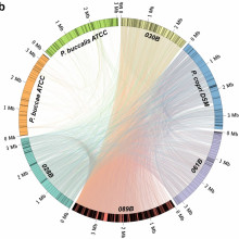
The gut microbiota of patients with rheumatoid arthritis is enriched in microbes belonging to the Prevotella genus. 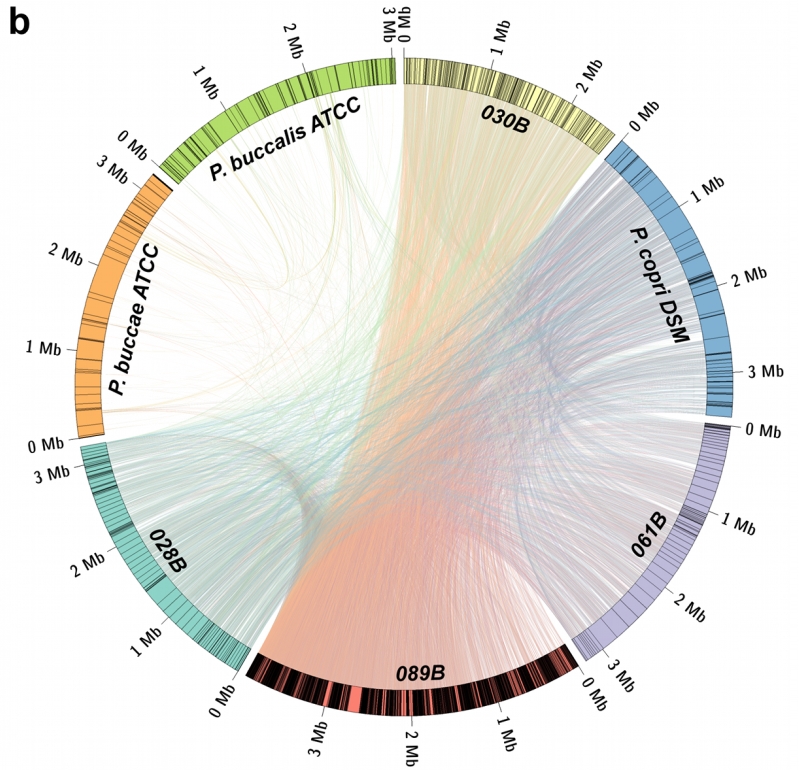
Dan - We found in mice, colonisation of the intestine with a particular bacterium thought normally to be a symbiotic harmless bacterium seem to trigger the onset of an arthritis syndrome that resembles rheumatoid arthritis (RA). And we were just asking whether we might see something similar in humans.
Chris - So, how did you actually approach this study? You took patients who had RA, rheumatoid arthritis, and then went looking to see if there were certain bugs that seem to keep cropping up in those patients.
Dan - Exactly. There's been a tremendous advance in the genomics of the consortia of bacteria that are present at various body surfaces. And advances in sequencing technology over the last five years have made it possible now to basically get a snap shot of the entire microbiome in one take. In our case, we were looking at the microbiota in the faeces and then get this kind of a sequence from each individual. What made our study successful, I believe, was that we had access to patients who had yet to receive any treatment. There were new onset patients who had had symptoms on average for only four to five months, and that's where we saw really the difference. And those people who had yet to be treated, we saw an enormous predisposition to be colonised with Prevotella copri.
Chris - So, this Prevotella organism, you find that it's over represented in this cohort of patients who have rheumatoid arthritis, in terms of its physical presence and the numbers of organisms that are there.
Dan - In terms of both numbers and presence, that's right. Prevotella copri seems to occur in a bimodal distribution in the population, particularly in western countries. And in the number of large scale studies, both in the US, the so-called human microbiome project, the HMP, and in Europe, there's another large study called the metaHIT study. In both of those, it was found that Prevotella is present in roughly 15% of healthy individuals.
Chris - So what do you think it's doing in these people who go on to get rheumatoid arthritis? What is the bug doing?
Dan - It's very difficult for us to know. Whether it goes along for the ride in somebody who's predisposed to rheumatoid arthritis and possibly, the inflammation in the body of these individuals favours the growth of the Prevotella. That's one possibility, but another possibility is the presence of this particular bacterium may actually favour the development of a particular type of inflammatory lymphocyte and that inflammatory lymphocyte may then go on to in some way attack the joints in the body.
Chris - We know also that rheumatoid arthritis runs in families. So, what role could genetics play here?
Dan - We know that there are particular genes that predispose getting rheumatoid arthritis. So, we looked at those particular genes to see if they were present or absent, and what we found to our surprise is that those who had the predisposition genes, actually had a lower level of colonisation with Prevotella. So, there was an inverse correlation. So, one way of interpreting this, is that there needs to be a particular threshold for activation of the immune system in order to trigger the autoimmune disease in rheumatoid arthritis. So, either you have the susceptibility genes, or you have a high level of Prevotella, but only one or the other is needed to trigger the disease.
Chris - So, what do you think it's going to take to nail this? How are we going to get to the bottom of the role that Prevotella might play?
Dan - Well, the important question now is to try to understand whether Prevotella triggers a type of immune response that can lead to increased inflammation in rheumatoid arthritis. And in order to do that, we need to do longitudinal studies in which we track patients who are at risk before they develop disease, and ask whether we can predict the onset of disease by changing their microbiota. In addition, we'll need to ask whether particular types of antibiotic therapies might be able to reduce the risk of disease. The other more mechanistic studies to try to understand whether there are particular types of immune cells that are specifically activated by Prevotella, because that will also give us a hint as to whether the Prevotella is acting through a direct activation of the immune system. And, related to that is the question of whether triggering is by a bacterium like Prevotella is sufficient to set in motion the disease process after which the Prevotella no longer has a role. Patients who have been treated long term, we don't see this increase in levels of Prevotella.
17:05 - Cortical plasticity: how brains adapt
Cortical plasticity: how brains adapt
with Tamar Makin, Oxford University
You can't teach an old dog new tricks, or so the dogma goes, and scientists say that this is because the brain becomes far less adaptable 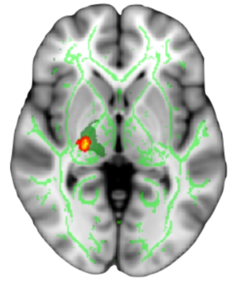 or plastic as we become older. But is this really true? Oxford University's Tamar Makin has been studying people lacking limbs to try to find out...
Tamar - Following limb loss, the major part of the brain that normally receives inputs and outputs to the hand is now deprived, and potentially, this could be very useful for people because perhaps we could tap into this deprived brain area and train it to support behaviour for other body parts, for example. And the question was whether that is possible and whether that opportunity for the brain to change would depend at the age at which individuals have lost their hands.
Chris - So, how did you do this? Who were you studying?
Tamar - We recruited several groups of individuals without a hand. One group is people that were just born without a hand, it was never fully developed, we call them congenitals. The other group is individuals that used to have a hand but it was chopped off due to traumatic circumstances, and these would be the acquired amputees. We had about 15 to 20 individuals in each group. On average, our participants were about 45 years old, but some of them were quite young, some of them were in their 20s, and others were in their late 50s.
Chris - So, good age range there.
Tamar - Yeah.
Chris - How did you actually study them?
Tamar - What we did is we put them in an MRI scanner and asked them to perform simple movements with different body parts. And using functional MRI, we were able to identify the brain areas that become active when they perform these range of movements.
Chris - And what do you see?
Tamar - We see that this deprived cortex specifically becomes activated either by the intact hand, that is the hand that hasn't been lost in the amputation and is still functioning, or the stump, so this is the residual arm or whatever was left from the arm after the amputation. And we see that the extent to which the deprived cortex becomes activated by movement depends not on the age in which amputees have lost their hand but rather to what extent they tend to use it in daily lives.
Chris - So, the more that they transfer the role of the lost hand to the intact hand, for example, then the greater the switch of allegiance of the patch of brain that was supplying their lost hand over to the now remaining hand then.
Tamar - Exactly.
Chris - Does that differ from what we thought might be going on before?
Tamar - Yes, in several ways. The important finding that we found in this study was that the deprived cortex is quite willingly being taken over. The intact hand is not a cortical neighbour of the missing hand. In fact, it's on the other side of the brain. And this is a very striking example of brain plasticity - of one brain area being reassigned or taken over to do something new. And the fact that this was more common in the acquired amputees, suggests that there's no upper limit in terms of age for plasticity, so that the brain is capable of changing even though people are quite adult and have had experience with that brain area and a particular body part for quite an extensive time.
Chris - How do you know - just playing devil's advocate for a minute - that the enhanced activation on the part of the brain corresponding to the lost hand isn't the reason they use the remaining body part more versus they use that body part more so that part of the brain develops to correspond to the movements in that body part more?
Tamar - So, this is actually a very good point because when we're using these tools, neuro-imaging, we can't pinpoint and say that it is the brain activation that is formed due to intense usage or rather individuals that have more plastic brains would therefore choose to use their intact hand more to make this inference, we actually have to follow people from the very first stage of losing their hand.
Chris - But, your inclination is that it is the use that begets the brain change, rather than the brain being capable of changing more which makes them use the surviving limb part more.
Tamar - Yes, and we also examined brain patterns of controlled participants. So, these are individuals with two hands and we were trying to see whether in just people that haven't lost their hand, activation in the hand area could be modulated by movement of the other hand. Let's say the dominant hand, when you look at the brain area of the non-dominant hand, and they all tended to show no activation when they were moving one hand in the cortex of the other hand. So, this leaves us to think that it is actually the behaviour that drives the plasticity and not the other way around.
Chris - Given what you found - what are the implications clinically for how we should rehabilitate people who are amputees or how we should help the people who are born with congenital limb loss?
Tamar - So, in terms of rehabilitation, we've always thought that there's a period in which the brain is more sensitive, is more capable of making changes, and that's the childhood. What our studies shows is that individuals have an opportunity to switch the way the brain is wired, and the way the brain is responding at every age. So, it doesn't matter if they lost their hand as children or were born without a hand, or lose it as adults, they should be able to use this deprived cortex to underlie adaptive behaviour.
23:37 - Why rodents have cone-shaped whiskers...
Why rodents have cone-shaped whiskers...
with Andrew Hires, Janelia Farm Research Campus, VA
Conical whiskers allow mammals that live in tunnels and other enclosed spaces to learn more about 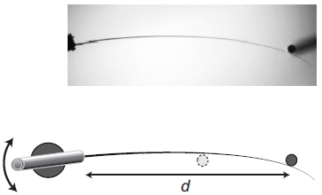 their surroundings.
Andrew - With mice, they have many whiskers and their sense of touch to their whiskers is vitally important for them to navigate their environment and to discover and identify objects around them. And these whiskers actually, have a very special shape. They're almost perfect cones, and this actually gives them a lot of advantages for the natural environment in which a mouse might find himself in.
Chris - So, what are the big unknowns then about these whiskers that you were trying to flush out?
Andrew - We really wanted to understand why is it that these whiskers are conically-shaped because all mammals have whiskers. There were other whiskers in other species that have different shapes like a flattened shape, or bulging shapes. So, what is special about the conical shape of the mouse whisker that is adaptive for the environment which mice live in?
Chris - Things like burrows, and holes, and sewers. So, how did you go about actually trying to model or test that?
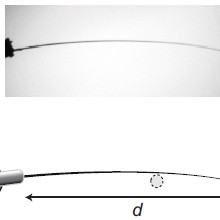
Andrew - As a scientist, the first thing you want to do is go the reductionist approach in which we take a whisker out of the mouse. Mount it on a little stick and then rotate it into objects at various distances. And when we observed how the whisker changed shape, based on how far away it is from an object, how hard we're pushing into the object. And what we observed is that when the conical whisker is moving into an object or being pushed past an object, there is a critical moment in which the whisker just slips past the object. It's not when the whisker reaches the very tip end. Actually, it's much sooner and we thought that that was kind of a surprise. And so, looking into the mathematics of how that took place, my collaborator David Golomb, realised that the precise point in which the whisker slips off an object is actually related to this conical shape.
Chris - Does it matter how far down the whisker that contact occurs before the slippage begins to occur?
Andrew - Absolutely. So, since whiskers are much more flexible at the tip than at the base, when you're contacting an object near the tip of the whisker, slip offs occur much more easily. You don't have to move the whisker very far before slip off occurs. Now, if contacting near the base, then you really have to rotate the whisker. The whisker has to move a long way into the object before it suddenly slips past.
Chris - So, do these measurements by physically mounting a whisker and then pushing it into things, is that actually what the mice do?
Andrew - So, what mice do is that it scamper up to an object and then they start moving their whiskers backwards and forwards very rapidly, then they orient their face so that they sweep their whiskers backwards and forwards across an object, that's called whisking. And over the course of moving their whiskers back and forth multiple times, they build up a sort of understanding of what the object is, its position, its texture, and use this to identify and localise the object.
Chris - So, do you think then that given what you've discovered about the importance of this conical profile, that we could use this in making better sensors for us rather than just for mice?
Andrew - This is just one interesting avenue of future research. If you look at any blind person that's walking down the street, often times they have these white canes that are going backwards and forwards, tapping left and right, left and right. And they can try to find out the distance to objects by striking them with their cane, and one critical thing I did when researching white cane travel, is I've read some people complain that, for example, a three-part white cane assembled in sections, lacks the same flexibility as a single uniform cane that has a taper to it. And I think that the same mechanisms are in play with blind people who are using white canes to navigate their environment and identify objects. So, it would be nice to see if we can now apply the principles that we've discovered from the mouse whisker system to improve the design of white canes, perhaps with tips that have a greater flexibility gradient from base to tip.
- Previous Life, The Universe and Everything
- Next AIDS Awareness Week


Comments
Add a comment