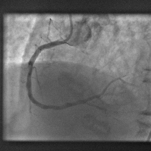
A dye used medically for over fifty years could be a shot in the arm for investigating coronary artery disease, scientists have found.
Writing in Science Translational Medicine, a team from Boston, US, led  by Harvard Scientist Farouc Jaffer have discovered that indocyanine green (ICG), chemical formula C43H47N2O6S2Na, which has been used safely for over five decades to measure blood flow through the heart and liver circulations, can be used to highlight inflammed atheromatous hotspots threatening trouble in narrowed arteries.
The team make the point in their paper that, although there are techniques for studying the progression of arterial disease in large calibre vessels like the aorta and iliac arteries, there is currently little on the table to aid in the identification of potentially troublesome areas for smaller arteries like the coronaries.
Seeking to address this challenge, the researchers surveyed a range of chemical tracers that might be helpful in identifying patches of rapidly progressive arterial disease. Such regions, which show more intense inflammation, are more likely to trigger thrombosis (clotting) and blockage of the vessel and are therefore a higher clinical priority for treatment.
Being highly lipophilic (fat-loving) the team wondered whether ICG might meet their needs and flag up fatty deposits in the vessel walls. It also absorbs and emits near-infrared light, making it a useful tracer molecule.
The team injected the substance into rabbits with damaged aortas, which are similar in size to human coronary arteries. Under a mciroscope, the ICG was found to bind with high selectivity to the diseased segments of the vessel. Moreover, it bound best in regions that were showing signs of rapid disease progression.
To determine how such an agent could be used for human imaging, the team inserted a probe into the aortas of a further group of animals and used near infrared imaging to plot the pattern of ICG signal coming from the inside of the vessel wall. This, they found, was a very close match with two other imaging techniques, one using ultrasound and another x-rays.
This shows that ICG could be used as a highly-effective and safe tracer to sniff out the smoking gun underlying arterial disease and therefore help cardiologists to direct their attention to those areas that need their input the most.
- Previous Attractive new magnetic material
- Next Electrons are spherical


Comments
Add a comment