A 3D breast in a dish?
Interview with
Jonathan, firstly, why are we so interested in modelling breast tissue?
Jonathan - Well, it's the most prevalent cancer in females. So, if you are able to model the normal situation, you are going a long way towards turning on oncogenes and introducing the tumour microenvironment into that normal situation, and actually, studying physiologically what happens in oncogenesis.
Kate - So, by modelling a breast tissue as it normally is, we can introduce  cancerous genes and cells and work out what happens and hopefully, from that point I suppose, work out drugs to try and stop that process?
Jonathan - That's right.
Kate - So, are you using human cells like Kelly is?
Jonathan - Well at the moment, we've produced a model that I'd say is physiological, to the point where it even produces milk. But we are now moving into producing a human model of breast tissue.
Kate - So, what's that model at the moment you're using instead of human tissue?
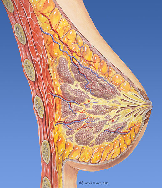
Jonathan - So, what we have is, a natural biomaterial which is effectively collagen and also proteoglycans. We introduce the principal components of a mammary gland into that model. First, the fat cells and also, the breast tissue itself which is the branching epithelium and we are effectively redeveloping the model in the dish in a natural form.
So, in the breast during puberty and in pregnancy, you get this massive elaboration of this tree and we're actually effectively doing that again in the dish. So, we're quite confident it's physiological.
Kate - You're putting these fat cells together, but you said it even lactates. Are there certain cells to do that or once you've put it in a model, does it automatically work out how to do that and form a breast tissue as a whole?
Jonathan - There's a certain amount of it automatically doing that, yes. You give them a little bit of nurturing and they'll seem to do what they do best.
Kate - Those fat cells that you're putting, do they come from humans or do they come from another sort of test animal?
Jonathan - As it stands, it's purely cell lines and these are derived from mouse.
Kate - How similar is breast tissue in mice and humans because if they're forming the structure that they used to, that they know about like you just mentioned, is that sort of physiological structure and layers, are they similar in both mice and humans?
Jonathan - They're relatively similar and obviously, the gland is a lot bigger as we know in the human. The compartment of different cells is slightly different. So, in a human, there are more fibroblast for instance.
Kate - Kelly just talked about having a pea-sized lung. If you're taking mice breast tissue, how small is that within our dish?
Jonathan - The scaffold that we use is actually, you cut it to any size. In terms of these small organoids formed within the scaffold, we're talking of about half a millimetre in length.
Kate - You're working towards building a model that replicates human that you can then introduce these cancer genes into. Once you get to that point, what can a 3D cell model tell us about breast cancer that other cancer research into genes or within sort of patients who have the disease can't tell us? What's it going to tell us that's different?
Jonathan - What's quite exciting is that we can actually move into looking at primary cells and these forms similar structures, but they form what's known as a basement membrane. So, they're enveloped by a particular array of proteins. What's exciting is that cancer is dangerous of course when it spreads and one of the things they have to do is they have to force themselves away or through this protein mix. So, we can actually monitor that in real time. You can't do that in any other way, so we're able to actually - because it's in the dish, you can actually see what's going on.
Kate - So, you can watch the cancer cells spreading along. Once we know more about that mechanism, what can we use that information for? Can we test new cancer drugs and see how that affects it?
Jonathan - Certainly, we can. The thing to realise about cancer, it's a highly individual disease. So, if we can form these small little mammary glands effectively in the dish and we can move into a human model, then we can test all manner of compounds on individual tissue from individual donors and that's very exciting because each person's cancer is different.
Kate - We're going to be talking in just a moment to some computer modellers who do their modelling theoretically. What advantages do you think that cell modelling has that if you looked at this from a computer point of view, you wouldn't be able to find out?
Jonathan - Computer modelling of course has its place, but the biological situation is much more unreliable and I think it was summed up by a talk I went to actually last week in London by a Professor of Oxford, Professor Phillip Maini who stated that in mathematics, if you divide 1 by 2, you get a half and in biology, if you divide 1 by 2, you get 2. It's basically unpredictable.
Kate - Them's fighting words! We'll see what Katherine and Peter have to say about that in a moment. Thank you very much to Jonathan Campbell from Cambridge University.


Comments
Add a comment