What is an X Ray?
First of all, how do x-rays work? Well, they don't, of course, any more than light 'works'. X-rays describe radiation which is part of the spectrum which includes visible light, gamma rays and cosmic radiation. X-rays were discovered by Wilhelm Roentgen, almost by accident, in 1895, and within a year the new technology was already in general use for imaging the human body. You can imagine how much excitement this development caused at the turn of the 20th century, not just for the medical profession, but also for the general public. It wasn't only respectable x-ray clinics that were being set up and advertised, there were also ads for demonstrations of the 'magic rays' at soirees, as a form of entertainment.
We consist largely of water
The point about x-rays is that, unlike visible light, they pass straight through stuff, including the stuff that people are made of. So, if you shine a beam of x-rays at someone, and put a piece of film on the other side of them, you produce a shadow of the inside of their body.
")
This only works at all because the different tissues that make up our bodies absorb x-rays to different extents. The problem is, though, that we consist largely of water, so most of our soft bits look much the same as far as x-rays are concerned. In fact, there are really only three tissues that are sufficiently different from each other in terms of x-ray absorption to show up on an x-ray film: bone, air and soft tissue. So, on a chest x-ray, for example (see picture, right), the bones (high absorption) are white, the air in the lungs (very low absorption) is black, and all the soft tissues (muscle, heart and blood vessels, skin) are a similar shade of grey. But even with that limitation, radiology developed into a major specialty during the 20th century.
Barium and other contrast agents
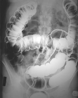
The problem of all soft tissues looking the same was solved to some extent by the introduction of artificial contrast material of various types. One of the first substances to be used in this way was barium sulphate. This is a salt of a heavy metal, and as such it absorbs x-rays very well. It can be made into a thick suspension and swallowed (the barium swallow or meal) or introduced into the colon via the rectum (the barium enema). The barium coats the lining of the stomach or colon, and when air is introduced to distend the gut, you get really detailed pictures of the bowel lining, covered in a thin layer of barium (the picture on the left) shows a barium enema - the barium is white, and the air distending the colon is black). Small tumours and ulcers can be diagnosed in this way.
Contrast media other than barium
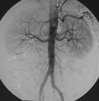
There are other contrast media, usually containing iodine, which are water soluble, and can be introduced into the body using other routes. For example, if you attach iodine to a chemical which is rapidly excreted in urine and then inject it into a vein, a series of films over the next twenty minutes or so will show the contrast material outlining the kidneys, ureters and bladder (the intravenous urogram, or IVU). A similar substance injected directly into an artery will demonstrate the vessel, and any organ (or tumour) which it supplies. This is known as angiography, and it is possible to pass a catheter (a fine plastic tube) from the femoral artery at the groin into almost any vessel in the body. The picture on the right shows the contrast material (black in this image) outlining the aorta, the main blood vessel carrying blood down through the abdomen to the pelvis and legs, and the renal artery supplying the right kidney. It was from this type of investigation that the sub-specialty of interventional radiology developed.
Interventional Radiology
What's the future of radiology?
These fairly straightforward methods for using x-rays have served us well throughout the 20th century, but from the 1970s on, we have seen the advent not only of a revolutionary (literally) new way of using x-rays, but also of several completely different imaging methods which don't use x-rays at all. As a result, you will fail to find an 'x-ray department' in many of our hospitals. Instead, there will be a 'medical imaging' department, the name change reflecting the wide range of technology now at our disposal. I'll talk about these new imaging methods next time...


 crystal")

Comments
Add a comment