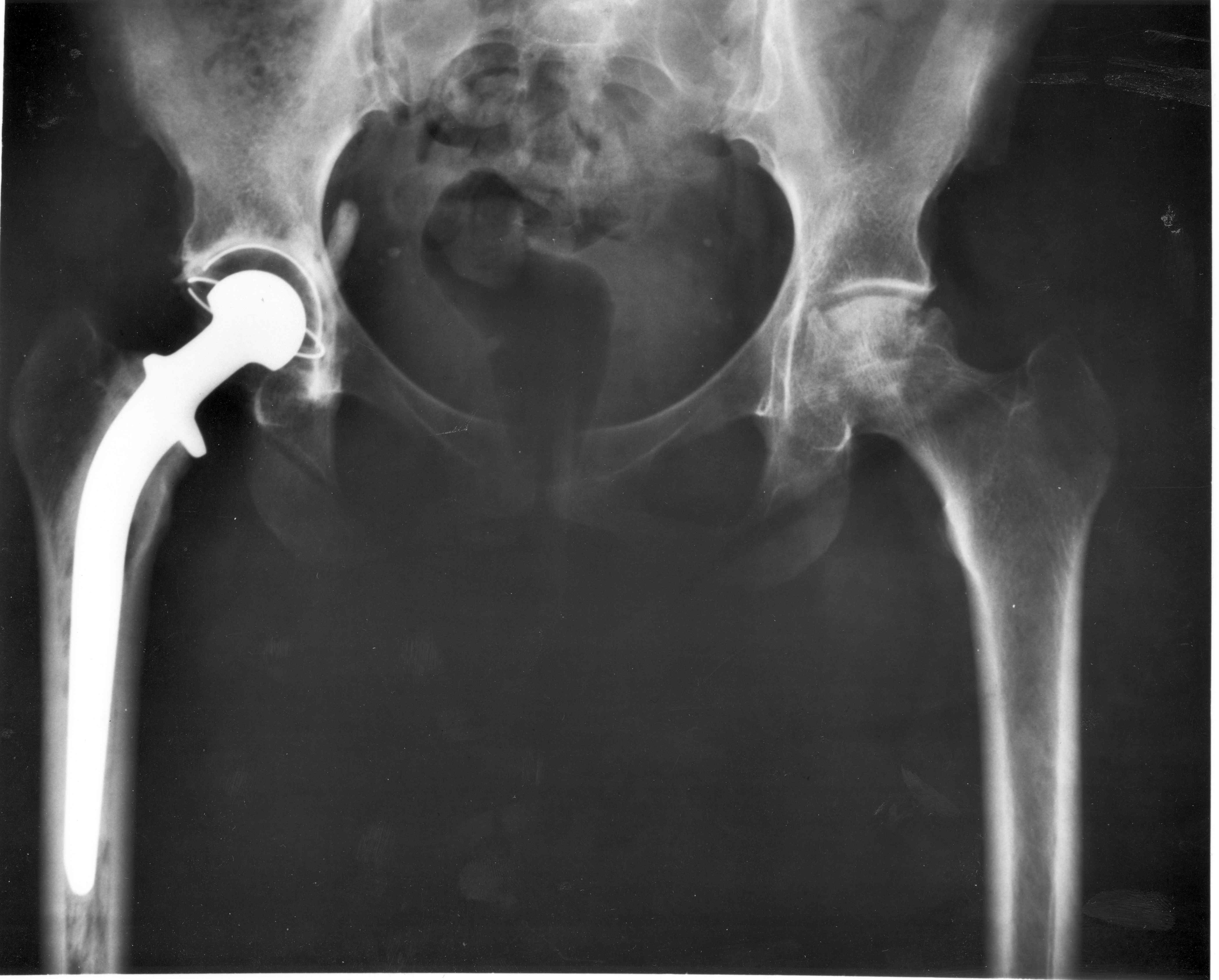
A new coating technique that makes bone bond tightly to an artificial joint has been  developed by scientists in the US.
Millions of joint replacements are performed each year, but in under a decade at least 12% of them will require surgical revision, half of these because the prosthesis has worked loose.
Currently, replacement joints are anchored in place using a bone cement compound called poly-methyl methacrylate (PMMA), which is disadvantageous for several reasons: most importantly, it has a different stiffness to native bone, so when the bone bends or flexes it moves a different amount to the cement, causing cracking and encouraging the two to part company.
Also, as PMMA is setting, it releases a large amount of heat, which can damage nearby tissue and also makes it difficult to include within the cement chemicals or growth factors that might help to promote local healing, or bone repair.
Efforts were made to produce porous prostheses intended to encourage bone to grow into the device, locking it in place, but these too have largely been abandoned owing to poor bone in-growth.
Now, step forward MIT's Paula Hammond. She and her team have instead developed a technique to apply a multi-layed, growth-factor impregnated coating to the surfaces of titanium or plastic prosthetic devices.
The base layer of the coating, which is about 1/5000th of a millimetre thick, contains hydroxyapatite (HAP), the very same calcium phosphate compound found naturally in bone.
Atop this HAP layer is a series dissolvable layers made from the bio-friendly polymers poly-beta-amino ester and polyacrylic acid. Impregnated into each of these layers is a growth signal called bone morphogenetic protein-2 (BMP2), which strongly stimulates the growth of new bone-building cells known as osteoblasts.
The thickness of these dissolvable layers, which build up like an onion skin and are formed by repeatedly dipping the prosthesis into a solution of the chemicals and then allowing it to dry, can be varied according to need. For a thicker layer, dip more; for a thinner layer, dip it fewer times.
Implanted into the shin bones of experimental animals, prostheses treated with the new coating showed steady release of the BMP-2 growth factor into the surrounding bone over about a 30 day period, and an ensuing in-growth of bone-building osteoblast cells.
Examining the implants under the microscope at time points up to 18 months later showed that the dissolved top layers had disappeared and new bone had grown up to and knitted itself into the HAP (hydroxyapatite) layer. There was no evidence of any tissue reaction to the implant.
Even more convincing was that, compared with untreated control implants, the force needed to remove from the bones prosthetics treated with the new coatings was over 30 times greater, and the test implants frequently came away with pieces of newly-grown bone still attached.
Writing in Science Translational Medicine where the work is published, the authors nonetheless acknowledge that they have worked only with rats so far and the technology needs to be tested in larger species and more realistic clinical settings.
But with the world's population ageing as it is, the demand for improved prosthetics with extended lifetimes has never been greater...
- Previous Keeping an eye on diabetes
- Next Maths maketh mortgage success


Comments
Add a comment