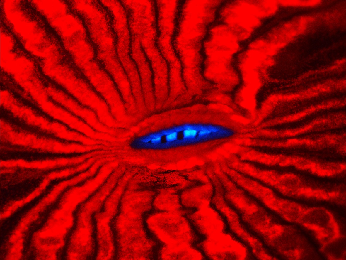
Scientists have reported in Nature Methods that a fluorescent protein called m-Iris-FP - originally isolated from the reef-building coral Lobophyllia - is ideal for high-resolution microscopy.
 |
Fluorescent proteins like GFP, originally isolated from a jellyfish species called the crystal jelly, have been in use since the mid-1990s. They allowed a massive breakthrough in the study of how cells work. Unlike other microscopy stains, that are highly toxic, GFP and similar proteins like this new m-Iris-FP can safely be used in living cells.
The gene that codes for the fluorescent protein is inserted into the genome of the cell or organism to be studied. When a particular protein is produced by the cell, the fluorescent protein is expressed as well and acts like a tag to track the protein's position and movement in cells and whole animals e.g. the distribution of a particular protein in the brain during embryonic development.
What's exciting about this new fluorescent marker protein, m-Iris-FP, is that it can be 'photo switched' to emit either red or green light.
The lead author, Jochen Fuchs, and his team showed that the marker allowed them to track dynamic processes in living cells at high resolution.
Obviously there is a LOT of computing power involved in this sort of microscope work, and the researchers had to genetically modify the protein to give it the exact properties they wanted. But it's pretty cool that life in the oceans continues to throw up new surprises and new avenues of improving research.
- Previous The proton just got smaller
- Next Cost effective coral restoration


Comments
Add a comment