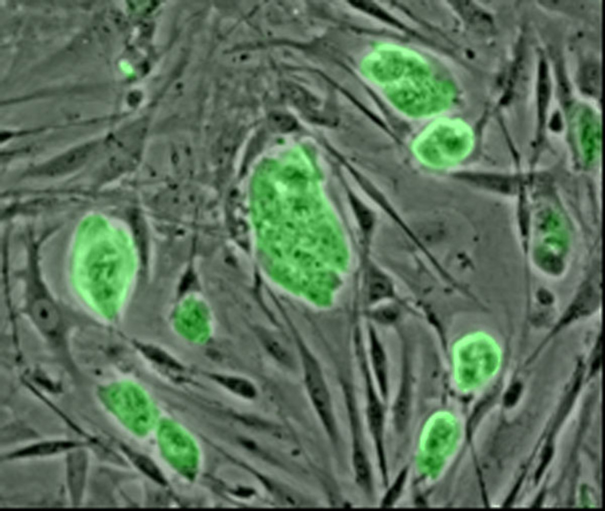
A way to label up stem cells so that they can be seen on ultrasound, MRI and microscope scans has been revealed by scientists in  the US.
As Sanjiv Gambhir and his colleagues point out in their paper in Science Translational Medicine, the promises of stem cell transplantation therapies for cardiac disease have yet to be realised, partly owing to the inability of doctors to identify appropriate areas for implantation and then follow up the outcomes for those cells.
To tackle this problem, Stanford University-based Gambhir and his colleagues have developed silica nanoparticles that are taken up by stem cells which subsequently become sustainably visible on ultrasound or MRI scans and will even glow up in ultraviolet light when specimens are imaged under a microscope.
So far they've tested the particles in the dish and in mice, but point out that they should be safe and well-tolerated in humans too.
As few as 70,000 cells labelled with the particles will show up and can be quantified under ultrasound, meaning that stem cells therapies can be objectively assessed to determine which clinical protocols, if any, offer the bets outcomes for patients.
- Previous Pushing forward with Planck
- Next Glasses-free 3D displays


Comments
Add a comment