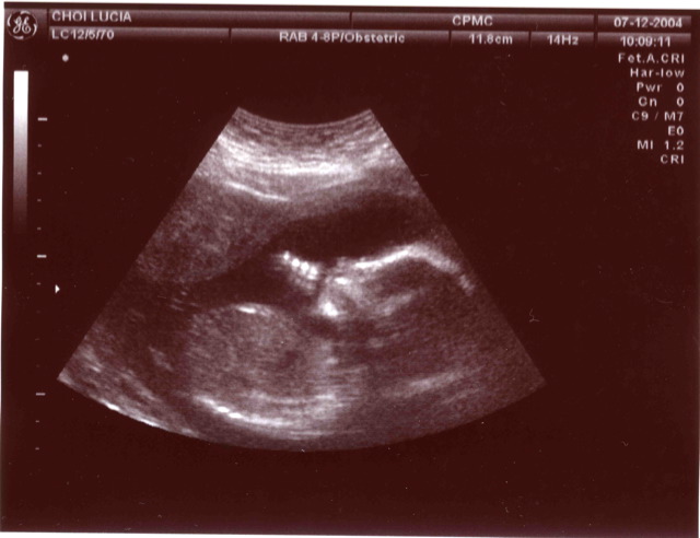
A way to read the entire genome of a developing foetus non-invasively and from as early as 5 weeks into development has been announced by researchers in the US.
Genetic defects, like Down's Syndrome, occur in about 1 in 500 births and these are becoming more common as women are leaving motherhood until later in life.
But diagnosing these conditions, and other rarer but equally devastating inherited disorders, requires a sample of the DNA of the developing baby.
The gold standard way to do this is with an amniocentesis or a chorionic villus sample (CVS) test.
Both involve passing a needle into the sac enclosing the developing baby in order to recover fluid or a small piece of the developing placenta from which foetal DNA can be extracted.
Between 0.1-1% of women who undergo these procedures may suffer a miscarriage, which is a risk many feel uncomfortable to take, and the earliest time at which testing can occur is from about 12 weeks.
Although progress has been made in recovering foetal DNA signatures that naturally spill over into the mother's bloodstream, this approach is hampered by the very low levels of foetal DNA, which also tend to exist as small fragments limiting the usefulness and reliability of the technique.
Now, writing in Science Translational Medicine, Detroit-based Sasha Drewlo, from Wayne State University School of Medicine, has developed a technique that can be used from just 5 weeks' of development and yields high-purity, intact foetal DNA and without any risk to the developing baby.
Drewlo's approach is to analyse the small numbers of foetal cells that naturally gather on the mother's cervix at the opening of the uterus.
These can be collected using the same technique used to obtain a cervical "pap" smear.
About one in a thousand of the cells brushed off is a foetal cell that forms a structure called the trophoblast, which is the tissue that forms the placenta and the membranes that surround a growing baby.
These foetal needles in the cervical haystack can be purified by mixing them with a special antibody that binds to a chemical marker called HLA-G. This is expressed exclusively on placental - but not maternal - tissue, so only foetal cells are labelled.
The antibody is also coupled to magnetic nanoparticles, so a magnet can then be used to pull out a pure sample of foetal cells. The intact DNA signature can then be decoded.
So far Drewlo and his team have tested the method on 20 pregnant volunteers. They compared the DNA sequences obtained using their new method with DNA extracted from small pieces of placental tissue taken from the same pregnancies.
Their Pap-smear samples, they found, provided the same sensitivity as the existing gold-standard amniocentesis and CVS tests but from just 5 weeks' gestation.
"We need to do more studies now to confirm that this works the way we think it does," says Drewlo. "But the potential is huge."


Comments
Add a comment