The brain is a hugely complex organ made from expansive networks of cells, all connecting and communicating with one another, quickly and effectively. This vast array of connections begin to form in utero, when the brain is rapidly growing and developing, and when the foetus is extremely sensitive to any kind of disturbance. In a paper published yesterday in the journal Science Translational Medicine, a team of scientists and doctors from the Wayne State University in Detroit outlined their ground-breaking work, developing a way to monitor the development of connections in the brain in the growing foetus.
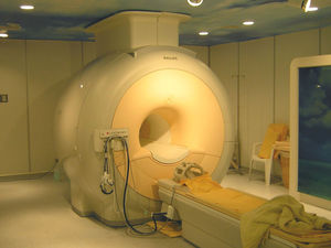
The team used a variation of the non-invasive MRI brain scanning technique to  assess connectivity in the brains of 25 foetuses. The technique, called resting-state functional magnetic resonance imaging, measures the magnetic differences between oxygenated and deoxygenated blood flowing through the brain. Active parts of the brain require more oxygen in the form of oxygenated haemoglobin in the blood, compared to inactive brain regions. The difference in oxygenated and deoxygenated blood can be detected by the MRI machine and can give us crucial information about where functional connections in the brain are being made, both at rest and during a variety of tasks.
The foetal scans demonstrate that brain connections are made during the second and third trimesters, between 24 and 38 weeks into pregnancy. Not only were the group able to successfully scan the moving foetus (not a small feat!) they were also able to show that connectivity gradually increases as the foetus grows. Scans at this stage of development have previously only been possible in premature babies, under situations where the brain may have been altered as a result of preterm birth. Of particular importance was the finding that many connections are made between the two sides of the brain, the cerebral hemispheres. Although the use of the technology in this way is in its infancy(!) the potential for effective and safe foetal monitoring is clearly a big step forward.
The value of these findings is emphasised by supporting evidence from other labs that show a variety of neuropsychiatric conditions, such as ADHD, schizophrenia and post-traumatic stress disorder, may be caused by alterations to the connectivity of the brain. As these connections appear to be developing in the foetal stages of life it may become possible, using this technique, to identify and correct the abnormal connections being made in the brains of patients suffering from conditions such as these.
- Previous Mars Simulated in Morocco
- Next Bone repairing gene therapy paste


Comments
Add a comment