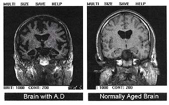
Scientists have developed a way to watch the pathological process that leads to Alzheimer's Disease as it occurs in the brain.
 Harvard researcher Bradley Hyman and his colleagues used a new imaging technique, called multiphoton microscopy, to peer into the brains of experimental mice that had been genetically pre-programmed to develop the rodent-equivalent of the disease.
This new approach meant that the team could look different distances into the same brain regions of single animals over a number of weeks to look out for the appearence of any abnormalities. Over the course of the study the researchers painstakingly matched up the locations of individual cells and blood vessels in each of the animals' brains as they looked for the development of amyloid plaques, the pathological hallmarks of Alzheimer's Disease.
These plaques are caused by a build up of a protein called beta-amyloid, which is naturally produced in the brain but is normally broken down and removed. Why it accumulates, how quickly and how this affects the brain surrounding tissue was not known, but scientists had concluded that the process probably occurs slowly, as evidenced by the pace of progression of the condition in humans.
But the new study has changed all that, because the researchers were surprised to see the plaques forming and evolving very rapidly. Amyloid deposits could appear in less that 24 hours in a brain region previously free from lesions. Interestingly, once the plaques reached full size they stopped growing possibly because other brain cells, called microglia, began to cluster around them. After this adjacent nerve cells began to show structural abnormalities, answering another existing question which was "which came first the abnormal nerve cells or the amyloid plaques?"
Although it's early days the new technique should provide researchers with a powerful new tool with which to probe the development of a disease which will inevitably affect one person in five in the developed world.


Comments
Add a comment