eLife Episode 59: Brain basis of blindsight
This month, the blind monkey that lacks a visual cortex but can still see, the bee-hunting wasps that use a gas cloud to keep harmful fungi at bay, adaptive optics that can image blood vessels of all sizes in the eye, the new field of palaeoshellomics, and how to mix a family with a scientific career...
In this episode
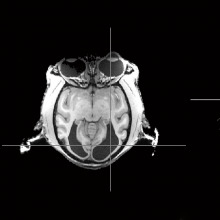
00:33 - The brain basis of blindsight
The brain basis of blindsight
Kristine Krug, University of Oxford
It’s often said that nature reveals her inner workings through her mistakes. What Kristine Krug discovered recently is no exception, and could give us new insights into what “blindsight” might really be, as she explains to Chris Smith…
Kristine - We studied the structure and the function of a monkey brain in an animal where a big part was naturally missing, and we looked at how the rest of it was working in order to support all the things the animal could still do which we didn't expect it to be able to.
Chris - How did you come upon this animal in the first place? Was it just by chance?
Kristine - It was actually a chance discovery. The animal was being trained for a different study, looking at big visual images and making decisions about it, and it had to just touch a big coloured object on the screen for a reward. And all her cage mates could do it, but that animal couldn't. And eventually we gave it a brain scan and found actually that the back third of the brain just wasn't there on both sides.
Chris - Why not?
Kristine - They think it was just probably born without this part of the brain.
Chris - And if you map the missing bits onto what we know about the structure of the monkey brain, which bits are missing?
Kristine -So it's at the back of the brain, which in monkeys and humans has basically a complete map of the visual world. All the information from the eyes comes in there and is then distributed further into other parts of the of the brain that does the processing for perception, and decision making, and behaviour.
Chris - We refer to that in a human as the primary visual cortex, don't we. It's the first jumping-off point for the visual information coming in from the brain, and we're comfortable that if we damage that - with a stroke for example - patients say they can't see anything once they lose that bit of brain. So how do you reconcile that with the fact that your monkey appeared to, at least some of the time, be behaving normally and be able to see?
Kristine - It's true, so in humans who lose that part of the brain late in life they are virtually blind, though they have some remaining vision called blindsight in some cases. So they can point to very bright moving spots of light even though they say they're not aware of them. And we think what happened in the monkey is probably an extension of this. There are other pathways that go from a relay in the middle of the brain to these higher areas, bypassing what is usually the major gateway. We think there was enough there for the rest of the brain to develop normally and getting a more extensive version of that blindsight which you can see in patients. And what was quite intriguing is when you offer normally a monkey a treat, the monkey will just come to the front of the cage and carefully look at the treat and reach for it and grasp it. That animal would start running past it as if to put the treat into motion and grab it out of the corner of its eyes.
Chris - One would infer from what you're saying then, that the monkey moved in order to make the object appear to be moving because then the motion decoding bits of the visual system that were intact in that animal could then register the presence of the object and provide information to the conscious brain so it knew what it was interacting with.
Kristine - Yeah. So all the behaviour pointed to that. And when we then put the animal in the scanner we could see that the network of brain areas that talk to each other when we or the monkeys see motion, they still talk to each other normally despite a major part of this network - the major input to it - missing.
Chris - Monkeys, like humans, are also heavily driven by faces aren't they. They are good at recognising other individuals and that goes on in the temporal lobe of the brain, a very different part of the brain. Was your monkey capable of recognising its colleagues?
Kristine - We don't know behaviourally, though the animal showed no problems with its cage mates. They interacted normally. But what we did do is we looked at these specialist bits in the brain that code for faces. And to be frank we didn't expect to find anything, because it needs very detailed fine vision for which we thought you need this primal visual cortex, a major gateway. But when you showed the animal faces of other monkeys, these very same face patches lit up in the temporal cortex as they did in typical monkeys. And we think that points to a separate input directly in these face areas from emotional centers in the brain being fed directly from the eyes.
Chris - Our existing view of how the visual system farms out the information that comes in is that everything ends up in the the first visual area of the back of the head and then from there it's parcellated off into these other centres that process things like colour, like movement, like faces. Is the finding from this monkey suggesting then that actually, that spreading out or that distribution of the information could be happening a lot sooner, a lot further upstream in the process, and that's why it's able to do this? Or do you think that this animal has just adapted to the fact that half its brain is missing, and so it's able to do these things because it's had to?
Kristine - We always knew there are small pathways that spread out earlier. So it is still the case that through the primary visual cortex 90-95% of visual information ends up in the brain network that supports our visual behaviour, but we knew about the other pathways. But we thought they weren't to that extent capable of supporting the rest of the network to function, and that's that's clearly been the case in that animal. We looked for strengthening of known connections of these smaller pathways and couldn't find any evidence, though we haven't looked at that exhaustively. But that's one avenue. It just could simply become...it's clearly not becoming stronger as a structure, these connections, but it seems like they become more efficient to drive the rest of the visual brain.
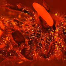
06:32 - Wasps use fumigation to keep food fresh
Wasps use fumigation to keep food fresh
Erhard Strohm, University of Regensburg
Some animals feed their young with breast milk. Some rely on their offspring fending for themselves. But others, like the wasp species Philanthus, also known as the European beewolf or bee-killer wasp, have a more gruesome strategy: they paralyse a bee, drag it to their lair and lay an egg on it. When it hatches, the wasp larva eats the corpse. We knew already that the wasps use several tactics to keep the food fresh, including bug-suppressing lipids and doses of friendly microbes, but now, as he explains to Chris Smith, Erhard Strohm has discovered they also use an even more ingenious strategy: fumigation…
Erhard - Insect eggs, actually the eggs of the European beewolf, fight off fungi using a gas. They have to do it because beewolves hunt honeybees and provide paralysed honeybees for their progeny as food. They bring these paralysed honeybees into underground nests, where there's a humid and warm microclimate. Because of this the bees will quickly be overgrown by fungi. And beewolves have already evolved two different mechanisms that we have reported on earlier: the bees are embalmed with hydrocarbons like lipids so that fungi grow less rapidly; the other thing is that beewolves have a symbiosis with bacteria, and these bacteria are cultivated into antennae and the females deliver these anterior to the larvae, and they integrate these bacteria into the cocoon where the bacteria produce antibiotics. But these two mechanisms were not enough to result in such a high survival of the larvae - so there must be a third mechanism. Actually it was the observation that when we open the observation cages we were reminded of chlorine of swimming pools. So we started to ask where this smell comes from, and it turned out that it was emanated from the eggs actually.
Chris - So you smell something reminiscent of swimming pools coming from these bees which have been embalmed by the wasp as future food for its larvae to eat. So where's that smell coming from then? The actual bee that's been embalmed?
Erhard - It's coming from the egg. You can just remove the egg from the bee and you find that the egg is smelling actually.
Chris - So the egg is releasing something, you could smell something coming from the egg reminiscent of a swimming pool: chlorine, but not necessarily chlorine. And it's that you thought that could be protecting the corpse of the bee?
Erhard - Yes. And we went on to identify this gas. We found that it was nitric oxide, and this nitric oxide is easily oxidised to nitrogen dioxide, and this nitrogen dioxide is what smells like chlorine.
Chris - And what does it do to microbes, that nitrogen dioxide? Does it kill them off?
Erhard - Well both nitric oxide and nitrogen dioxide are both radicals, and they will react with biomolecules and they will kill the fungi.
Chris - Why don't they kill the wasp larva then inside the egg? And also why don't they wipe out the good microbiota that the female wasp has wiped off of her antennae and impregnated onto the corpse of the bee?
Erhard - Yes, this is one of the most puzzling things about this whole story. We don't actually know how the egg is protected. We think the embryo in the egg produces this NO, and this NO might be transported to the eggshell with special transport molecules, and then it's delivered outside, but it can't go back into the egg. So maybe it's just a layer outside or on the outside of the egg that prevents these nitrogen oxides coming back into the egg. And the other question, why the bacteria that are delivered by the beewolf female into the blood cell are not affected, also is not known yet. We can speculate that these bacteria have evolved a resistance against these nitrogen oxides but we don't know yet.
Chris - Given the advantage to the fungus is so huge, that there's all this wonderful bee food to eat if you're not being poisoned by nitric oxide, why hasn't the fungus evolved to be more resilient in the same way that you're presuming that the microbiota which are being added to the bee by the female wasp, and the larva growing inside the egg; why has the fungus not developed its own resistance?
Erhard - Well if there would be selection for resistance in the fungus, they certainly would have evolved a mechanism to withstand the nitric oxide or nitrogen dioxide, but there isn't huge selection on the side of the fungus because in all brood cells there would be different fungi. And because of this non specialisation there is no selection. The symbiotic bacteria are always exposed to the nitrogen oxides, so there will be strong selection on them to produce resistance. The same with the embryo of course.
Chris - And do the larvae shut down this overproduction of NO as their gestation develops?
Erhard - Well a very interesting aspect is that the gas is only produced during a short period of about two hours and it starts about 14 hours after egg laying, and the gas will be produced for two hours and then it fades away. So it seems that there is one time point of huge concentration of this gas, and during this time the fungi will be killed and then the gas will disappear from the brood cell, so the larvae then they hatch will not be affected anymore by the gas.
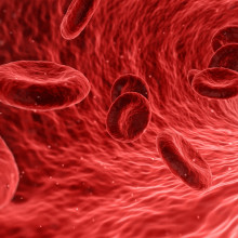
13:19 - Seeing blood vessels in the eye
Seeing blood vessels in the eye
Aby Joseph, University of Rochester
When we gaze skyward at night, stars appear to twinkle because the light path passes through patches of warmer and colder air, and this bends the light rays. Astronomers use a technique called adaptive optics to detect and correct for these aberrations and still produce sharp images. And now scientists in the US have used the same trick to image non-invasively and in detail the blood vessels - and the movements of individual blood cells - through the retina. And some of what we thought we knew about blood flow in the eye turns out to need another look. From the University of Rochester, and speaking with Chris Smith, Aby Joseph…
Aby - What we have here is a new method to non-invasively visualise and measure single blood cells flowing in the smallest to largest vessels in the retina. If you look at the brain, one of the most metabolically active organs in the body, we've gotten a lot of insights into the functioning of the brain using functional MRI. However there are still some limitations to that; one of them being that the resolution such techniques can go to is about one millimetre, and it may miss changes happening at scales much lower than that, namely in the smallest vessels of the central nervous system. So what we've done here is we've borrowed a technique which was first pioneered in astronomy called adaptive optics. Our view of the stars above in the heavens gets distorted and blurred because of turbulence in the atmosphere, and mirrors which can change their shape on the ground adaptively correct for that turbulence and sharpen our view of the stars in the sky. And we've borrowed this technique to image the retina.
Chris - So how do you get from what's going on in the night sky to what's actually going on in the back of the eye?
Aby - Imperfections in the front of the eye cause the image to be blurred. So we use adaptive optics to get a high resolution of cells in a living eye. Additionally we used a very fast camera to now start visualising blood cells which obviously are moving, and combined this with the computational technique to now be able to measure speeds of blood cells from the smallest capillary to the largest vessels in the eye, giving us a complete picture of the interconnected network which becomes important in studying disease.
Chris - It sounds amazing. So talk me through then actually what the technique is. Because you're doing this initially on test animals aren't you? You're looking at mice to start with. But talk us through then what you actually do and how you obtain the images.
Aby - What we do is we use an infrared light source which is non-invasive and to which the eye is relatively insensitive. We scan a very fast beam across blood vessels in the eye and we're able to image back the light that is scattered by the red blood cells. And because we are imaging fast enough we're able to measure speeds of up to one metre per second in the eye, covering the entire possible range of speeds of blood cells from the smallest to the largest vessels.
Chris - So you have a light source sitting outside the eye which is beaming near-infrared through the pupil and the optics at the front of the eye. It's hitting the blood vessels which are actually behind the retina, bouncing back off those, and some of the reflected light comes back out the front of the eye where you can see it. And you're doing that by scanning in a series of lines a bit like a television would scan across the screen, in order to get a complete picture, many many times a second.
Aby - That's absolutely right, yes. 15,000 lines per second enables us to not miss anything that's happening.
Chris - How do you know that you're seeing the same bit of eye each time? Because obviously the eye might move a bit, your apparatus might vibrate a bit, and at the resolution you're looking - fractions of a millimetre - any tiny movement would blur things. So how do you match them up?
Aby - That's a great question. So it's partly done by the adaptive optics technique which adaptively corrects for changing imperfections at about 10 times a second. And additionally in post-processing we have what we call registration or correction algorithms which can correct for the eye drift or eye motion.
Chris - And so this gives you the architecture of the vessels, it gives you where the cells are going in those vessels, so that you can image right through from the smallest vessels to some of the largest in the retina. And that doesn't just mean the blood coming in, you can also therefore look at the veins that are taking the blood away as well presumably?
Aby - Exactly, yes. And that's what we think is one of the advances in this paper, where we can look at the complete vascular unit, which becomes important for example in diseases like diabetes where it's known that the disease may start at the smallest level of the capillary and have a cascading effect to larger and larger vessels. And this technique now enables us to study the complete system.
Chris - Have you got any surprises emerging from this? Because physiologists have been studying how cells move along blood vessels for a really long time, and we've got various theories about what happens when blood flows through different sizes of blood vessels. When you actually now look at them rather than just go by what the theories say, were there any obvious differences?
Aby - That's a great question. And perhaps that's one of the unique selling points here. So what we find is, contrary to models which predict a certain shape to how the blood distribution is in a cross section of a vessel, we found that when we actually directly imaged them or visualised them that it's quite removed from what conventional models predict of how blood flow should look like at the smallest vessels. And this requires more study, but some of the first results indicate that we indeed need to directly measure some of these metrics, and not only rely on models because there can be a lot of surprises when it comes to these extremely tiny vessels.
Chris - Indeed but also big vessels. Because we're doing things like building stents to open up blocked arteries for example, and the way these are designed is based on our ideas about how blood flows through blood vessels, and how blood cells interact with the walls of blood vessels. And actually if those models are a bit wrong it argues that we may not be designing these instruments to go inside blood vessels as well as they could be designed.
Aby - Yes, certainly. And I couldn't agree more, and one of the advantages here is that although we're looking at the eye and the retina, because the retina is a part of the larger central nervous system which is a very special tissue with a very dedicated vascular network, what this really enables us to do is study what's happening in the nervous system - as opposed to the skin which is more easily accessible - and see if there are differences in these models or assumptions, as you mentioned, of what's happening in blood vessels in the nervous system.
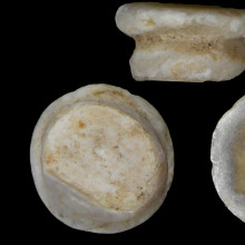
20:23 - Palaeoshellomics - proteins from old shells
Palaeoshellomics - proteins from old shells
Beatrice Demarchi, University of Turin
An Italian team are taking modern science and applying it to the past, both to understand biology better and to solve an important archaeological riddle. From the University of Turin, speaking with Chris Smith, Beatrice Demarchi...
Beatrice - The first question we had was we wanted to know if we could apply new molecular tools to invertebrate calcified tissues such as shells. We really know very little about the biochemical evolution of these organisms. But at the same time we also had a very specific archaeological question from some colleagues in Denmark: they asked me whether I could identify the material, the shell that had been used, to make some special ornaments called double-buttons. Everyone thought that the shell that they used was an oyster, because at that time in Europe - and we're talking about 6,000 years ago - there were different cultures and different ways of making ornaments, and understanding which materials they were using would give us an understanding of the view that they had of the world.
Chris - And you couldn't get that information from anything else? The context and so on wouldn't tell you that?
Beatrice - Actually interestingly the context led us astray if you will, because this was a coastal site, a shell midden of oyster shells. And so we all thought that this was a sensible material to use for these ornaments, and of course that turned out not to be the case. And morphology couldn't help us anymore because these ornaments - they're really really tiny, they've been worked really heavily, in this case they were worked to resemble pearls. And so all the morphological features, they're just gone.
Chris - How did you resolve that then? You've got something where there's not enough morphology to tell you what sort of shell it's come from, you just know it's come from some kind of mollusc or something. So how did you take it forward?
Beatrice - That's right. We only knew that it was shell, and only after we really looked at it with a microscope. We used a series of different techniques that could tell us broadly whether these shells were marine or freshwater, and that was important. The same technique also told us that these shells were likely to have been collected locally. Then we looked at the microstructure using electron microscopy. And then finally we had to develop a whole new strategy based on molecular analysis: we looked at proteins. Proteins in shells are very special; they can survive for thousands or even millions of years, but we don't know much about the sort of sequences that the shells have.
Chris - Now when you say the sequences, do you mean as in what the proteins are actually made of, the building blocks, the amino acids that are in there, and therefore what the gene sequences that the organism would have used to assemble them in the first place? Because obviously if we know what the protein's made of we have some idea as to what the organism was, because we can identify almost genetically.
Beatrice - That's right, yes. By looking at these sequences we hope to get some through genetic information. The problem is that we know very little about the DNA sequences even of mollusc shells. We only have about 15, 20 genomes of mollusc shells; a phylum that has something like 100,000 species.
Chris - I was going to ask you that! Because yes you can get the protein out, yes you can potentially - assuming it's not too degraded - work out what the sequence of the amino acids is in that; but without a database, a reference database of all of these proteins in all the different molluscs and shellfish and shelled creatures, you're not really much further forward are you. So how how did you get that database?
Beatrice - That's right. We were actually stuck with it for a few years. And then we were very lucky because a transcriptome of a freshwater mollusc was released and suddenly our sequences made sense. After a couple of years of just having sequences without any reference we could map them on some sort of scaffold. And it was very clear that our main proteins were freshwater-mussel-type-of shell proteins, and so we could take the work forward. But the other thing that we did do was to build our own reference dataset. So we looked at modern and archaeological shells from the same sites and we had to develop some fancy bioinformatics approaches for that.
Chris - Serendipity is a wonderful thing in science, isn't it, when it happens like that! What does that therefore tell you about the archaeological question? Because once you managed to get this needle in a haystack and you knew, right, this is a kind of freshwater mussel, does that move you forward?
Beatrice - Well we couldn't really pinpoint them geographically, but the big surprise was that these freshwater molluscs, they were found lonely in this shell midden made of millions of marine shells. So we were on a winner in that sense, that suddenly we had a whole new perspective on our archaeological question. Because in general our expectation is that to make prestigious objects you need prestigious materials, and in Central European sites where these double-buttons are quite common, people used... well we thought that people would have wanted to use exotic shells, so marine shells for example from from the Mediterranean, but our coastal Danish site was the exact opposite, we had freshwater shell probably coming locally. And so this was telling us that wherever we were in Europe, people had a very precise idea on how to make these double-buttons and they didn't really care about whether the material was exotic or not in that sense. They cared about the actual properties of this material. Mother-of-pearl is a beautiful material, it's really hard, it's really resistant, but it's also really easy to work. And so this is giving us a whole new insight really on how people were thinking about materials around their environment.

27:05 - Mixing science and a family
Mixing science and a family
Kathleen Flint Ehm, Stoney Brook University
Each month on the eLife Podcast we also try to consider some of the social aspects of science and life as a scientist. This time, we’re looking at the scientific workforce, which has changed dramatically in recent decades. For a start there are far more women progressing through the discipline. But the scientific career structure itself has not kept pace with these changes, particularly when it comes to the question of fitting in a family. Kathleen Flint Ehm is the Director for Graduate and Postdoctoral Development at Stoney Brook University. Talking to Chris Smith, she explains how she's spent a lot of time looking at this issue across her career…
Kathleen - I hear stories from postdocs who are doing job interviews from the delivery room. I hear about researchers who are coming back from maternity leave and doing work far before their doctors would recommend. And so I think somewhere in between the fear that someone is going to take a year off of research because of pregnancy, and the reality of these early career moms who are barely taking time for themselves to recover before they turn back again to their research and their career; somewhere in between there something has to give. I think part of that is that we need to change the culture around childbearing and family-friendly environment.
Chris - So what do you think it is that's got to give? Do you think the funders should exert influence and say, well rather than have this time-bound funding, that means automatically the project's going to suffer because you're going to run out of road if that person takes a year off; or do you think the institutions have to say, well we'll make the institution more friendly; or actually is it both?
Kathleen - You know that's a really good question, and I think the simple answer is that it's both at some level. Now funding agencies work differently in different countries and have different types of constraints. Here in the US our federal agencies that fund most of science have actually gone on the record as being quite family-friendly, and have put a number of policies into place that have been trying to make research more amenable to taking a short break. For example, providing funding for temporary technicians to come into the lab to do work while a key team member might be out on leave for whatever reason. And I think that these are a great start, but I think the environment and frankly personnel policy is really set by the institution. And so in practice you need to change the local environment and the local personnel policies at the individual institutions.
Chris - The other issue is of course science is an international pursuit, and many people bolster their CV by working all over the place. And that's great but it's not family-friendly either.
Kathleen - No it's not, and this was actually something that we found in some of the focus groups that we conducted when I was at the National Postdoctoral Association. One of the big issues that comes up of course is: for every woman who's going to start a family, start having kids, adopting children, having that broader family support network is very important. For academics who are rolling wherever the next job may be, it's harder to have those local networks. For international women it's even harder. And so that was a concern that I heard over and over again of, do I want to stay here in this country? Do I want to move back to my country and be nearer to my family? And I think once upon a time... we really think about who the typical researcher was, and the way that for example the postdoctoral phase of research was designed, it was really designed around the people who were doing science a couple of decades ago. They tended to be single men who could easily move around at the drop of a hat for any new position. And scientifically I still hear a lot of people talk about the importance of moving from institution to institution in order to broaden the reach of your science, to have more diverse ideas, and overall to be able to increase innovation.
Chris - Do you think the situation will solve itself in a way? Because the generation of people coming through tend to be of a different mindset than people of yesteryear. But also we have new technology coming to bear, and it's becoming a lot easier for instance to work from home productively. You don't necessarily hold yourself back through spending a bit more time not in the lab.
Kathleen - The danger is that if you're constantly in touch you also never have that distance from your science. But I I think that in this day and age we have some cultural shifts just naturally in the way that we do work. But I think that at the same time science is also becoming inherently more collaborative, and you're having these larger and larger teams. If it's science that requires big instruments or big labs or things like that, some of it still needs to be done in person. So I think that we still need to be mindful of these issues even as the models for doing science are also evolving.
Chris - So if you had a young woman who's an early career scientist sitting in front of you now, what would be your number one, number two, and number three most important pieces of advice you would give her, for both a successful but a family-fulfilling career?
Kathleen - I think my very first piece of advice would be to sit back and take stock of the things that you find most satisfying and successful for success, and to define what success means for you. I think my next step would be: really think about what success or satisfaction also means for you in your personal life, whatever that may be. That may include family, that may not, but understanding your own personal values is just as important as understanding your technical skills and where you want to go with your job. My third piece of advice in particular for those early career researchers who are thinking about starting families is: make sure that you have a supportive partner. Half of, I think, the great advice that we hear from people is that for all the good planning that you do, no plan is perfect; and so being able to have a team on your side that can help you get where you want to go and balance all your priorities is going to be really key.


Comments
Add a comment