This week we're in for a little light relief, as we explore how light-based technologies are delivering a brighter future, in medicine and beyond. Plus, in the news, a new gene therapy for spinal muscular atrophy, scientists make sonic holograms, and the most accurate reconstruction of a dinosaur yet.
In this episode

01:07 - Genetic test for heart disease
Genetic test for heart disease
with Professor Nilesh Samani,The University of Leicester and The Bristish Heart Foundation (BHF)
A new genetic test to diagnose individuals at risk of coronary  heart disease at a much earlier stage has been unveiled. An international team of scientists married up over 49,000 inherited genetic markers called "snips" - single nucleotide polymorphisms - with whether or not 15,000 individuals carrying those markers had developed heart disease. This has enabled them to identify a pattern of markers that together can spell out the likelihood of a person going on to have heart disease later in life. And this means that it might be possible to intervene and ward off a heart attack before it even happens. Nilesh Samani is one of the study authors and he took Chris Smith throughtthe details.
Nilesh - Heart disease is the commonest cause of early death in this country and in many countries round the world. There are many reasons - important lifestyle factors: smoking, lack of physical activity, having certain conditions such as high blood pressure and diabetes, high cholesterol, but there is also a very important genetic component to this. Perhaps about fifty per cent of the risk of getting heart disease is related to an inherited component.
Chris - And so is that what you're probing with this present piece of research - trying to get a handle on how those genes play a role in the development of the disease and finding out who's got them?
Nelesh - In the last few years we spent a lot of time trying to identify genetic differences between people which affect the risk of getting coronary heart disease - we've found quite a few of these now. But this particular research takes us beyond that point and asks the question, if we had this genetic information on individuals, how well does it predict them getting heart disease?
Chris - If we know we already have these risk factors, are we not already pretty good at identifying who the people are who are likely to suffer a heart attack?
Nilesh - No because of two reasons: the risk factors like high blood pressure or cholesterol only explains part of the risk of getting heart disease and, to a large extent, the genetic factors that we have identified are independent of those. Some of them work through these risk factors but the majority of them don't so they provide independent information. What we know is that coronary heart disease, the underlying process, starts in people in their twenties and at that stage the risk factors we currently use are not particularly discriminatory between individuals. Whereas your genetic risk factors - you're born with them and so we can look at them at any stage in one's life and, therefore, put people into different categories of what their genetic risk is.
Chris - And how did you do that?
Nilesh - So we created from the studies we've done where we've identified the genetic variations, we've created a genetic risk score. And then, on the basis of that risk score, we look to see whether those individuals who had the highest burden of genetic risk (top 20 per cent), versus those who had the lowest burden, what their lifetime risk of getting coronary heart disease was in a number of populations that we had access to around the world. And what you see is that if you are unlucky as an individual to have the top twenty per cent of genetic risk, then over the lifetime your risk of getting coronary heart disease was at least fivefold higher.
Chris - Armed with that information, how do you propose that we use it?
Nilesh - Obviously more work needs to be done to show that this finding can be applied to different populations under different situations. But if we confirm that then I think that one of the things that we can do is to identify those individuals who are at highest risk much earlier, at a time when the process is developing silently and to try and prevent it progressing by giving them advice about lifestyle. Perhaps giving them some treatments, such as statins, at that very early stage in the disease process rather than waiting for the condition to develop.
Chris - Although the study doesn't tell you whether that intervention will work, does it? Because what it does say is you can identify people who carry these high risk genes but it doesn't tell you if we intervene in those people whether we can affect the outcome?
Nilesh - You're absolutely right. We have some evidence that they probably do because in some other studies investigators have shown that if you carry a high genetic risk burden and you give statins to those individuals, versus people who carry a low genetic risk burden, then the absolute benefit from the statins is much higher in those who had high genetic risk. So we know that if you can identify those people even some of the treatments we have now are likely to be of benefit. But you are absolutely right that we need to do more studies to both demonstrate that having this knowledge will be be beneficial. And secondly that this is something that the public will accept in terms of screening and finally that it is cost effective for health to be able to supply it.

05:40 - Sound holograms
Sound holograms
with Dr Andrew Mark, The Max Planck Institute for Intelligent Systems
Take a close look at your bank card or passport, and you will probably see a  hologram, used to make the document harder to fake. The surface of the hologram is minutely textured, embossed with tiny bumps that are the code for an image. When light waves bounce off the surface, they reconstruct the image so that it appears to float in mid air. Now, researchers in Germany are applying the same principle with sound waves. They 3D-print a piece of plastic to create a sonic "lens" that shapes sound waves emerging from an underwater speaker so that they form an acoustic "landscape". This could be used to move cells around in a dish or even to treat diseases inside the human body. Laura Brooks heard how it works from inventor Andrew Mark...
Andrew - What we're trying to achieve is controlled shaping of the sound field. We'd like to have particular areas that are high intensity and other areas that are low intensity, and we do that through what we call an "acoustic hologram." The idea is that we have a transducer and a transducer is basically a speaker. It projects a sound wave through the hologram and the hologram acts effectively like a lens to modify that sound wave so that in the far field or downstream of the hologram we have a well defined shaped sound field.
Laura - So can we think of it as something like a landscape that has hills and valleys of sound, effectively?
Andrew - That's exactly right. So the hologram itself is a piece of 3D printed plastic and it has tomography to it. So certain pixels within the hologram are higher or taller than others and because the speed of sound within the hologram is different than within the water in which it's immersed, the waves have different phase when they leave the holographic plate.
Laura - And what kinds of patterns are you making and what would you use them for?
Andrew - So one of the things that we try to do is create a two dimensional image downstream from the hologram and we can shape that image into the shape of a dove, for instance. And we use that to do particle collection so we have many, many small microscopic particles that collect into the sound field into the shape of the dove.
Laura - So you're drawing pictures then with these sound landscapes?
Andrew - Exactly right!
Laura - That's really impressive. What else can you do with them?
Andrew - So one of the other things that we can do is we can change the arrangement of the hologram to project sound upwards towards the surface of the water. So the hologram is under water and it projects sound waves up to where the water meets the air. And where there's higher sound intensity we get crests forming in the surface of the water and, if you put particles onto the crests, then the particles are trapped there. So one of the nice things that we can do is shape the profile of these crests so that they don't just have simple points but they're instead linear tracks for instance.
And the other nice thing is, because we have such complexity in this hologram, we can actually build a phase gradient into the track and the phase gradient serves to actually push the particles. So now we have a track that the particles are confined to and a phase gradient that pushed them along the track. We can make these tracks into shapes like, for instance, letters or we can do rings and we can have multiple rings. And, in some cases, we can have particles that move in opposite directions depending on the direction of the phase gradient along these rings.
Laura - So you can actually move things around without even touching them - just using these sound fields?
Andrew - Exactly.
Laura - So you've talked about assembling these tiny particles but what could this be used for in terms of practical applications?
Andrew - One of the things we have in mind is to use it for either therapeutic or diagnostic medical energy. So one of the applications that ultrasound is used for right now is for either ablation or for thermotherapy where it's used to heat things deep inside the human body.
We can imagine a scenario where a doctor takes an image of the patient's body - a particular patient. Figures out what the best way or the best distribution of sound field inside the patient's body is, graphs a hologram by 3D printing that is specific to that patient, and then uses that hologram to heat or ablate in a way that's particular to that patient's needs.

09:54 - Does sugar make kids hyper?
Does sugar make kids hyper?
with Dr Kat Arney, The Naked Scientists
In this week's Mythconception Kat's been sifting through the sweet stuff...
Kat - That's right! Picture the scene - a kids' birthday party, complete with a table laden with sugar-packed goodies. Just a few slices of cake or choccie biccies seems to turn the mildest child into a hyperactive monster, quickly creating a situation of knee-high near-anarchy. But while it's a commonly-held belief that dosing kids up on sugar makes them hyperactive and badly-behaved, like a badly-organised pile of party cookies, the scientific evidence behind these claims just doesn't stack up.
Brave scientists have actually carried out research into the links between children's diet and their behaviour, dating back more than 20 years. For example, in 1994 scientists published a major study in the New England Journal of Medicine describing a nine-week-long, placebo-controlled trial looking at the effects of sucrose (that's regular table sugar to you and me) and the artificial sweetener aspartame on the behaviour of nearly 50 pre-school and school-age kids - including children whose parents reported them as being particularly sensitive to the hyper effects of sugar. What's more, they specifically picked children whose parents said they were sensitive to sugar. Although it's difficult to carry out these kind of studies with 100 per cent accuracy - kids will be kids, after all, and it's hard to completely control what they eat and do - and although the studies usually only involve a relatively small number of participants, the researchers concluded that "neither sucrose nor aspartame produces discernible cognitive or behavioral effects in normal preschool children or in school-age children believed to be sensitive to sugar."
This finding was backed up by another paper published in 1995 - something called a meta-analysis, which combines many studies looking at a particular question to see if there are any effects once a larger number of participants is grouped together. Again, it concluded that "sugar does not affect the behavior or cognitive performance of children. However, a small effect of sugar or effects on subsets of children cannot be ruled out."
So given that the studies say that there's either no effect, or maybe only a tiny one, why do parents still swear that sugar turns their little angels into little monsters? It actually seems to come down to parental expectations. Put simply - if parents see their children eating lots of sugary food, they believe that they're going to act up, and so attribute any naughtiness to the sugar. This was even tested in a placebo-controlled trial with mothers and sons, where half the mums were told their children were necking a sugary drink, while the others were told they were getting a drink with artificial sweetener, even though they were all given the artificial sweetener rather than sugar. But the mothers who thought their sons had drunk lots of sugar were more likely to say their child was acting up. The mums themselves actually criticised the apparent sugar-drinking kids more, and watched them much more closely.
In fact sugary drinks seem to alter parents' behaviour rather than children's - if you believe sugar is going to make your child behave badly, that's what you're going to be looking out for. And it goes without saying that children's parties are ground zero for reinforcing this idea - lots of sugary foods on offer, plus over-excited kids doing lots of fun activities that get them hyped up, and an expectation from parents that it will be a living nightmare. But it's more likely to be that expectation - along with the overall excitement of the event - that is responsible for unruly behaviour, whether real or imagined.
There's still research going on to find out whether sugar - or, indeed, artificial sweeteners - have any link to childhood conditions such as ADHD, but for now there isn't enough hard evidence to be sure. But of course, nobody is saying that it's fine to let kids guzzle on sugary drinks, cakes and chocolate bars all day long. After all, healthy foods such as fruit and vegetables all contain certain amounts of sugars in various forms. But there are plenty of good reasons for kids - and adults - to keep an eye on the amount of sugar in their diet. For example, eating lots and lots of sugary foods increases the likelihood of obesity, which can cause problems later in life. But bad behaviour doesn't seem to be one of them.
14:04 - Spinal gene therapy
Spinal gene therapy
with Dr Suzan Hammond, The University of Oxford
A gene therapy breakthrough for an inherited motorneurone condition called 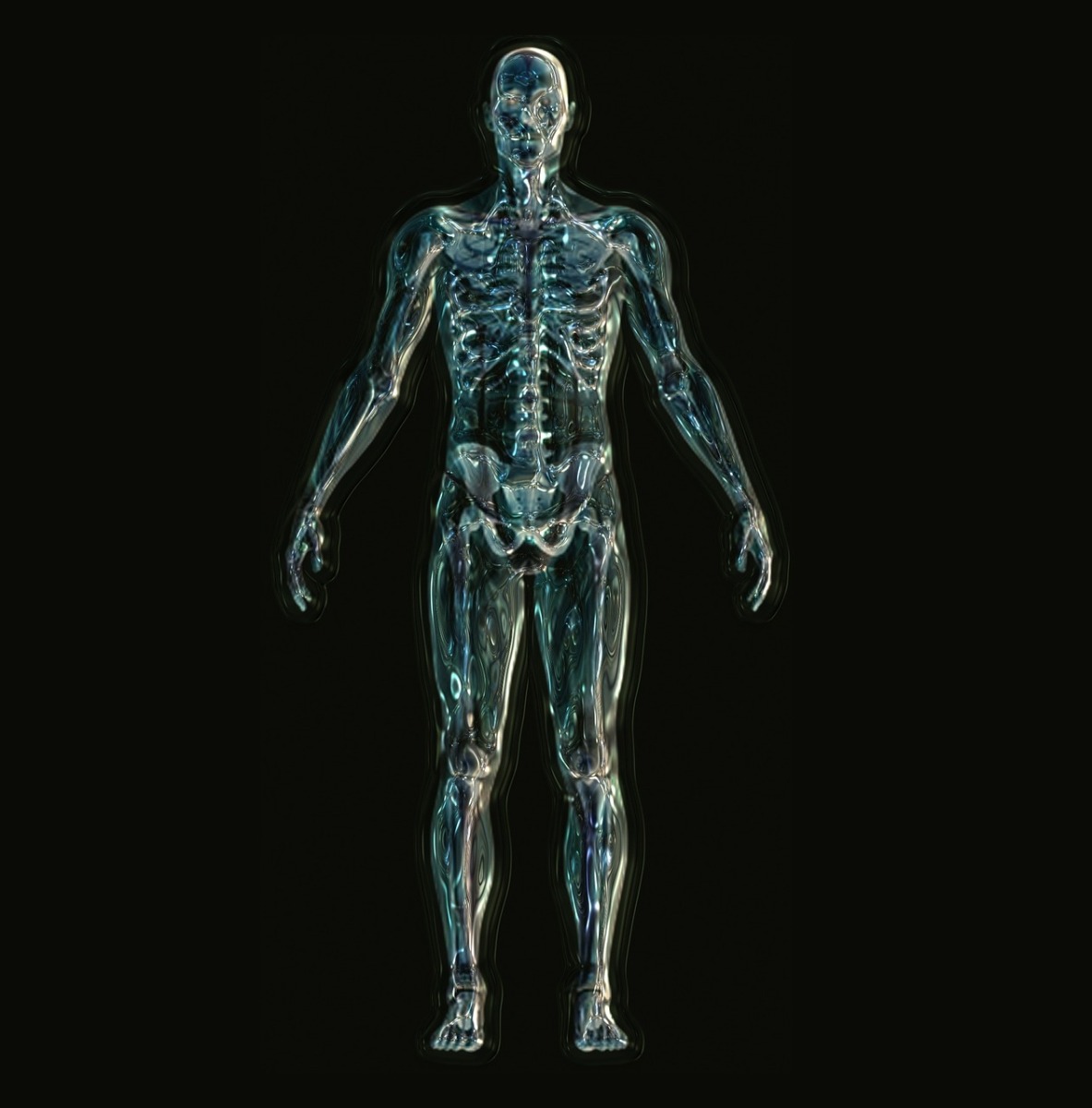 spinal muscular atrophy, or SMA, has been unveiled by scientists at Oxford University. Patients with SMA cannot make sufficient amounts of a protein called SMN - short for "survival motorneurone" - which is coded for by a gene called SMN-1. What Suzan Hammond and her colleagues are announcing this week is a DNA sticking plaster or "patch" that can boost the activity of a backup gene, called SMN-2, that can make up for the missing SMN-1. The way they get this genetic patch into the affected cells is by coupling it to a short protein, or peptide. Chris Smith found out how the technique restored a normal lifespan to mice with the animal equivalent of spinal muscular atrophy...
Suzan - Spinal muscular atrophy is an inherited disorder that mainly affects children and starts to present between zero and six months of age. It results in death in the motoneurons in the spinal cord and these motor neurons they reach out into the skeletal muscles. So when they die the skeletal muscle becomes disused and it starts to become degenerated. It's caused by a mutation in the survival motor neuron gene - the gene that's inherited for creating SMA.
Chris - Given that this is a genetic disorder - how might we be able to tackle it then?
Suzan - So, in SMA, we have two forms of the gene that causes SMA - SNM-1 and SNM-2. And in most of us SNM-1 is fully functional and will give all the protein that is needed to survive and to be healthy but in patients there are mutations or deletions in SMA-1. So we left with SMN-2, which only gives about ten per cent of this protein that you need which isn't enough for survival. So what we try to do is we try to enhance the amount of protein that SMN-2 gives.
Chris - So there are two genes (SMN-1 and SMN-2). A healthy person who doesn't have this condition has a normally working copy of both but SMN-1 produces the vast bulk of the protein that you need from that gene. And in people who have damaged that gene, they get this condition but they do still have the SMN-2 gene limping along and you're saying - can we pep that up in some way in order to bring them up to the level that would make them healthy?
Suzan - Yes. So we're quite lucky to have the SMN-2 gene that every patient has so with one kind of treatment we can target on almost one hundred per cent of patients.
Chris - Right, So how do you try to persuade SMN-2 to rev up its production so that it produces enough of the stuff you need to keep the cells healthy?
Suzan - So, in SMN-2, it creates a product which isn't what we would call "functional." And what we have designed is these short bits of DNA, which act as a patch, in which it can combine itself very well onto these products and, therefore, make it a functional product.
Chris - How do you get those patches into the cells that need them?
Suzan - On their own it's very hard to get these patches into the cells. We have designed something which we call "peptides" which you could think of as a passport or a key that you add to these patches and that really allows these to get into the cell that is required.
Chris - How do they work - how do they do that?
Suzan - It's a little bit unclear. It's very non specific - it's the type of charge that these peptides have and it seems to allow uptake of these patches into the cell.
Chris - So you have a lump of protein - these peptides which you couple to the patch so one's holding hands with the other. And the passport - the peptide gets it into cell and then the patch - the short sequence of DNA does the business once it's in the cell?
Suzan - Yes, exactly.
Chris - And does it go everywhere in the body or does it target the delivery to those motor nerve cells that are most vulnerable in this condition?
Suzan - It goes everywhere in the body.
Chris - So how do you know this works - what tests have you done to show that this has got legs?
Suzan - We've treated mouse models of SMA that are very, very severe, so these models will only survive till about twelve days after birth. And when we introduce these peptide patches into these mice on the day that they're born, they increase their survival to a mean of around four hundred and fifty six days.
Chris - From what - a daily dose or once?
Suzan - Twice - it the day of birth and then two days after.
Chris - Good grief! So two doses is enough almost to give these mice a normal life span because mice don't live that long do they?
Suzan - Yes, it's essentially a normal life span.
Chris - And it goes into the bloodstream?
Suzan - Yes.
Chris - What about the function in these mice? I know you're saying that they have a normal life span but do they appear to be functionally normal - do they get muscle problems or do they appear not to?
Suzan - They're essentially normal. They're as strong as normal mice; they're bigger than untreated animals and their what we call "neuromuscular junctions," which is the area where the nerve hits the muscle - that's completely normal as well.
Chris - And do you think that this protein - the peptide passport - will also work in humans?
Suzan - well we certainly hope so. If you inject pups on the day that they're born we say they have sort of less developed biological barriers so it would be a bit easier for this to get through the body. But we can do it in the adult animals and, in the adult animals, we still see it getting into the brain and the spinal cord. And so we really hope this will translate into humans.
Chris - And so, the next step?
Suzan - Well the next step is to try to make this a clinical trial. We are working to get this through all the things that need to happen to put it into a clinical trial - the safety data and that sort of thing. And then, in a couple of years, we hope to start recruiting for a clinical trial in this.
19:59 - How the dino got its shading
How the dino got its shading
with Dr Jacob Vinther, The University of Bristol
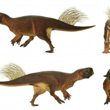
Scientists from the University of Bristol have revealed the most accurate  reconstruction of a dinosaur ever made. A fantastically preserved fossil, found in a lake deposit in China has revealed the colouring of the creature, but also where it lived before it found itself at the bottom of the lake. Georgia Mills spoke to researcher Jakob Vinther from the University of Bristol, to get to know this cretaceous character..
Jakob - Well the dinosaur is called "psittacosaurus" which means the parrot lizard. The derivation of the name comes from the fact that it's got a beak that kind of looks like a parrot's beak. So this is a distant relative to triceratops and it's about the size of a labrador, and it's from some deposits in northeastern China in the province called Liaoning that we find these fossils in, which is about one hundred and twenty million years old.
Georgia - I looked at some of the reconstructions of this dinosaur from your paper and it's quite cute. It's got these little sort of cheek horns that stick out at the sides and it looks like quite a friendly vegetarian.
Jakob - Yes, this dinosaur was definitely extremely cute. Now that we finally have a reconstruction of what this dinosaur looked like we can see that it's exorbitantly cute. It kind of looks like somewhere between like maybe E.T. and then I don't know what else you can compare it with. It kind of like looks like E.T. - it's got a very cute, wide face but then it has these very wide cheek horns that make it look very, very distinct.
Georgia - And what have you been investigating for this dinosaur?
Jakob - We have been looking at a fossil of this psittacosaurus which is extremely well preserved. What we have is remains of the skin associated with the skeleton and psittacosaurus is a fairly common fossil in these deposits in northeastern China. But occasionally we get some of these specimens that got washed out into these lake deposits and this one is really, really complete and really, really well preserved.
So we have looked at these skin impressions and we find organic residues in these. And we've recently discovered when we have organic remains, for example, feathers preserved in dinosaurs, we had the pigments retained in these and we can look at these pigments, which is melanin, and make some inferences about the original colours and colour patterns that this dinosaur would have had. So now we've done the same thing with this dinosaur, we have looked at these organic residues and when we look at them under a so-called scanning electron microscope, then we find little structures which are melanosomes. So that demonstrates what we have preserved in this fossil is the melanin pigment that gave the colour to the animal.
Georgia - OK. So you've actually found the fossilised remains of pigments and that has then, in turn, informed you of what this dinosaur's colouring looked like. So what can this tell you about the dinosaur other than helping you having these lovely reconstructed paintings of it?
Jakob - So that's the interesting thing - we can also say something about what this dinosaur was doing because it had these colour patterns for a reason and these colour patterns that we see in this dinosaur are various types of camouflage patterns. And the most important one, which is the one we best can use to say something about the environment that it lived in is this transition in light to dark between the back, and the belly, and the tail of the animal, which is this pattern called "countershading."
Georgia - So, putting everything together, and we've got a labrador sized dinosaur, it's got a body like a kind of chubby t-rex, and the head looks confusingly like a cross between ETs and a parrot. Also there's some weird grass looking stuff sticking out of it's tail. And then the colouring is this speckled black and red on a tan body, which is much darker on the top and this is what Jakob referred to as countershading.
Countershading is a clever technique that's still used by animals today. It involves having a lighter underside and a darker back. Think about things like penguins, deer, rabbits - they all have lighter bellies and this basically makes you look a lot less conspicuous. Our brains are trained to spot objects when they are lighter on the top and darker on the bottom as that's how the sun tends to light things up. But, if you counteract this with your shading - countershading - you tend to appear more flat and this makes it easier to avoid being eaten as predators have a harder time spotting you.
So this dinosaur seems to have been using this trick over a hundred million years ago and this also tells us something about its environment.
Jakob - Because what we see in living animals is that the countershading varies depending on the habitat that they live in. And the reason why is because the light conditions will vary depending on where you live. So the countershading in animals that live in savannas, for example, are usually sitting very high on the body and they're very sharp. Whereas animals that live in a forest, they have a countershading which is much more gradual and it sits much further down on the body.
So, basically, it means that we can use this sort of evidence to go back to reconstruct this dinosaur and put the original colour patterns onto it and we can show that it lived in a habitat which was a forest.
26:03 - WaterScope - preventing disease
WaterScope - preventing disease
with Dr Richard Bowman, The University of Cambridge
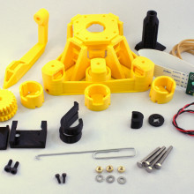
Contaminated drinking water is a major source of disease in poor countries.  Thousands of children die every day from waterborne illnesses. Fortunately, Cambridge scientist Richard Bowman is working on a low-cost solution called "WaterScope" a hand-held plastic microscope that can test water samples for infectious disease.
Chris - Richard - how did this project get started?
Richard - Well, it got started about a year ago now, when a student project looking into how we can do water testing faster and more cheaply, came up with the idea of using a microscope to essentially shrink existing tests and do them much faster. What the microscope does is tell us whether particular water is clean or dirty. Normally you do this by collecting some water and incubating it overnight with some nutrients for the bacteria, then the following day you can see colonies of bacteria with the naked eye, and that's how you tell if the water's clean or dirty.
If we do the same experiment under a microscope, you can see the bacteria after more like an hour and so you can get an answer much more quickly.
Chris - Now that's very laudable but, of course, people in poor countries can't afford expensive, high-end microscopes which is, presumably, why you're going down the route you are?
Richard - Absolutely. We want to make the microscopes very cheap, very easy to use and, ideally, we want to be able to make them in the countries where they're needed to avoid having to ship stuff in from the UK, which is very expensive.
Chris - But, if you are making the case that a poor person can't afford an expensive microscope, can they afford an expensive 3D printer?
Richard - Well, we don't need every person that has to test their water to have a 3D printer, but we're starting to see labs setting up, where they do have 3D printers and they're looking at ways they can use these to produce useful products like, for example, our microscope.
Chris - So how do you 3D print a microscope and are they any good?
Richard - They actually work really, surprisingly well. The trick that we've used to make the microscope is quite an old one. Essentially we take the wide angle lens that you find on most webcams and turn it upside down, put it slightly further away from the camera sensor, and it becomes really quite a good little microscope objective.
The other ingredient is holding your sample in the right place and being able to focus it. And we do that by, essentially, instead of sliding stuff around, which is hard to do on roughly made 3D printed components, we use the flex in the plastic to actually bend the structure to move the sample into focus and move around on it.
Chris - That's very clever. So how do you actually build one of these things - just talk me through piece by piece - is it like airfix? You know those kits where you used to make little aeroplanes from pressing out the things - some of the them about the size of a proton from what I can tell(!) and assemble them into a working gadget.
Andrew - Yes, it is a bit like. The microscope prints in I think it's ten or eleven pieces, depending on exactly what you build. Most of it actually prints in a single piece so all of the sensitive flexure mechanism that handles focussing it and translating the sample comes off in a oner. Then you have to add in three screws, which is what you use to move the stage around and to focus the microscope, and snap in the camera and the optics. So the whole thing - well it takes me about twenty minutes-half an hour but I've built quite a lot of them now.
Chris - But even so - that's not long is it? And the proof is in the pudding though - does it work? If you take a sample of dirty water from a puddle or a river and put it under the microscope - can you see whether or not there are germ-forming bacteria there?
Richard - Absolutely we can. We're still working on some of the sample collection stuff, so going from the dirty water to something on a microscope slide that we can image. But the imaging part works really well and the rest of it we hope to have sorted out in the next couple of months.
Chris - And once it is sorted out - how does it get deployed and where are you targeting?
Richard - In the first instance we'll do a few small trials with a couple of partners. Take it out to places where they're already doing water quality monitoring and test our system versus theirs. Once we've established that it's all working nicely, we would work with the same partners to start getting testing kits out with their aid workers so they're being used by someone that will feed back to us how they're working and how we can make them better.
Chris - And, can you also effectively establish a surveillance network by having lots of these on the ground in lots of countries afflicted by waterborne illness problems, does this give you data that you can then share in order to anticipate need for healthcare and also tell people there's a problem coming - don't drink the water?
Richard - Yes, exactly. And one of the most powerful things about it is that it is all built around a computer - in this case a Raspberry Pi. So all the test results are digital and we can send them back and build a database and sort of map out where the problem is.

31:24 - Visualising tumours
Visualising tumours
with Dr Sarah Bohndiek, The University of Cambridge
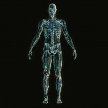
Sarah Bohndiek's Vision Lab at the University of Cambridge is using light to fight cancer. She joined Kat Arney to show how...
Kat - So tell us a bit about this - how important is imaging - being able to see stuff, to fighting cancer, to diagnosing it and to treating it?
Sarah - Well imaging is very important in the journey of a cancer patient. So if you think about the development of the disease early on, many of the symptoms of cancer are nonspecific. So patients will often go into their GP and report these nonspecific symptoms, who will refer them into the hospital.
Now if we didn't have imaging techniques like X-rays, or Magnetic Resonance Imaging (MRI), then we'd have to surgically open up the patient and search for the tumour mass.
Kat - Oh my God!
Sarah - So having these non-invasive imaging techniques allows us to non-invasively have a look inside the patient and search for any abnormal lumps bumps which shouldn't be there in the normal anatomy. So it's very important at diagnosis but then it's also very important when we come to treatment, for example.
So if we think about setting up radiotherapy. Many solid tumours are treated with doses of radiation which are shaped and formed to target the tumour mass, and if we weren't able to see noninvasively where that mass was, then we might misdirect our dose and could cause side effects to off-target tissues.
So it's important in diagnosis, in treatment, and then many patients undergo follow-ups so they'll come back routinely to the hospital to have scans over time to check that their tumour is shrinking, and so that's how we manage to monitor that the tumour is responding to any therapy that's been given.
Kat - So you've mentioned a couple of techniques - a CT which uses X-ray, MRI which uses magnetism. But what are you doing - you're using a different form of waves aren't you?
Sarah - That's right. So we're trying to use visible light and light that's just outside that visible region. And the reason for that is that CT and MRI and other conventional imaging technologies that we're familiar with in hospitals are relatively expensive and require patients actually to go into the hospital and have a scan that's operated by a specialist person. If we could make more cost-effective technologies that could be distributed to GP surgeries, for example, then we might be able to actually improve the journey of a cancer patient where they can get a faster diagnosis rather than having to go into the hospital.
Kat - So, you've got a little demo here to demonstrate one of the techniques that you're using. Talk me through it - what's the technique and show us how this actually works?
Sarah - So the technique is called "photoacoustic imaging" and it's based on a physical concept called the "photoacoustic effect," and this effect is quite readily understandable. If you think about going out into the sunshine on a hot day, the light energy that the Sun is beaming down will be converted into heat energy in our skin, so light is often converted into heat. So that's the photo part of the photoacoustic effect.
So the acoustic part is the heat being converted into sound. So the advantage of this technique is that we can measure how light is absorbed in tissue but we can image it using sound which penetrates much more deeply into the tissue. I mean, we know we can't see through each other so that means that visible light doesn't penetrate very deep into the tissue, but sound does. And so, using the combination of light energy to create sound, means that we can get much better images.
Kat - so you've got a nice little demo here - How does this work?
Sarah - So the demo that I've got is a Coke Zero can..
Kat - Other sugary drinks are available - non-sugary drinks!
Sarah - Thanks Kat. So the reason for using this is the can has a black coating on it and black is very strongly absorbing of light. You can do this at home with any regular camera flash that you have in your house. You can put it close to the coke can... so I'm now going to put the camera flash right up close to the coke can.
Kat - About how far away would you say that is?
Sarah - So about a centimetre from the coke can.
Kat - OK. Flash right up against the can...
Sarah - And I'm going to flash it... and you hear a little 'ping' of the coke can.
Kat - Wow! It's like someone's kind of flicked it with a finger.
Sarah - Exactly. So I'll just do it again so everyone can hear it...
'Ping'
Kat - Wow!. OK, so that is the light basically being turned into sound?
Sarah - Yes. So that's the energy coming from that camera flash being turned into a 'ping' of sound. And so that's exactly what we're doing with the photoacoustic effect when we use it for imaging cancer. So we send a 'ping' of light in; In this case we use a pulse of laser light, and that pulse of laser light gets absorbed and it produces a sound wave which we can detect using regular ultrasound transducers.The same systems that you would use when you're imaging a foetus during pregnancy, and we can use those to detect the sound.
Kat - So you're listening to the light coming back at you?
Sarah - Exactly. And so the advantage of doing this is that ultrasound penetrates much deeper into the tissue than light does, but light gives us much more better contrasts. When we do ultrasound imaging we're just looking at reflection of sound off of boundaries so we're not looking at any particular kind of molecular information or any functional information.
Whereas when we use light, light is absorbed by a number of different molecules in the body, including haemoglobin in our blood cells. And haemoglobin has a different absorption spectrum so it absorbs light differently with different colours depending on whether it's bound to oxygen or not. So that means, in a tumour, we would be able to develop a map of the level of blood that's present in the tumour, so how many blood vessels are feeding it and also, how well oxygenated that blood is, so how well the nutrients are being delivered.

36:57 - The First Response in an emergency
The First Response in an emergency
with James Baker, Cambridge Design Partnership
Light-based sensors can give us important information about a patient's condition - such as their heart rate, or how much oxygen is in their blood. Laura Brooks has been to see how a new light-based device could help save lives in an emergency...
James - This is James Baker. I'm a partner at Cambridge Design Partnership and I look after electronics and connected products.
Laura - This week, Cambridge Design Partnership (CDP) received two awards for developing a wearable medical device that could help medics save lives on the battlefield.
James - At the point of injury, the battlefield medic faces a very challenging situation. They're trying to rescue somebody, they're trying to keep them alive, and there's many distractions, there's probably significant danger. And the process that they go through is to look at breathing and heart rate and base the kind of care that they give off those readings. But measuring those parameters is quite challenging and whilst they're measuring them they're not providing care.
Laura - A challenging situation indeed! But CDPs First Response Monitor could support medics in their difficult task. The gadget uses light based sensors, among other techniques to measure a casualty's vital signs...
James - A little piece of focus technology that can give them those important parameters and let them concentrate on providing care brings real benefit and helps them to improve the casualty outcome.
Laura - The little monitor is only about the size of the top joint of my thumb. It has a small display screen on the front and a clip on the back - a bit like a clip on an earring except that instead of clipping onto your earlobe, it clips onto your nose...
James - It needs to clip onto the nose because we're interested in measuring both heart rate and breathing rate. The heart rate can be measured using an optical technique but the breathing rate we do by looking at changes in temperature and humidity that are influenced as you breath in and out.
Laura - Can you talk me through how it works?
James - Yes. We shine light into the tissue of the nose and that gets bounced around and diffused and reflected inside the tissue and some of that gets reflected back to the device. And looking at the amount of light that gets reflected back, it's influenced by the amount of blood that's in the tissue of the nose, so that gives you a signal that's related to the heart rate with blood being pumped into the nose and then moving out again.
Laura - Is that similar to how fitness trackers work that people might have at home?
James - It's exactly the same technique, so on many fitness trackers you'll see a light on the back that's contacting the skin and its using exactly the same technique to measure heartrate.
Laura - That's neat, I always wondered how they work. So what else can you sense with this device?
James - Staying with the optical technique, if we actually use two slightly different colours of light shone into the tissue of the nose, we can look at the fact that blood cells change colour dependent on how much oxygen they're carrying. So looking at the ratios of those two light wavelengths that come back to us, we can actually determine the oxygenation state of the blood.
Laura - So you record all of this data from the patient - what do you then do with that data?
James - There's multiple levels of how we use and display that information. The primary method of display is that those three parameters are shown on the device itself - there's a nice little bright OLED display on the device.
On the next level we can actually communicate that information off to a wireless device, such as a mobile phone or a tablet and that allows us to do two things; we can show for a single casualty how those parameters have changed or trended over time, but it also allows us to monitor multiple casualties so you can start thinking about which one of my patients needs my attention most urgently.
Laura - You've got your smartphone here - can you show me how it works?
James - Certainly! So you can see on this front page we're currently connected to one device and we're showing those three parameters of respiration, heart rate, and SPO2 for that one device. We could have multiple devices connected to that front page.
Or, alternatively, you can dive more deeply into the information from any one device - so I'm pressing that one - and it's brought up some graphs that are looking at those instantaneous parameters but it's also showing me graphs of how those parameters have changed over time.
So you can see, for example, the breathing graph had me breathing at about twenty two breaths per minute but now you can see where I've taken the device off and it's now trended down to zero breaths per minute. I think if the device was still attached, that would be a big alert for you to actually go and have a look more closely at me.
Laura - James told me that this kind of monitoring also answers another key challenge in treating casualties in dangerous situations - how to keep information about the patient with the patient while they are transported to safety for further treatment. Since the wearable monitor stays with the casualty the data that it records is automatically passed on to rescuers and is available to whoever is caring for them next. Once more, James thinks the monitor could give lifesaving assistance in a range of different scenarios...
James - We originally started by investigating military uses but we see lots of opportunities for this in all sorts of civilian first response situations as well, such as disaster relief or anywhere there's a multiple casualty situation.
It can bring real benefit where you have first responders - people on the scene - who have some level of medical skill but they're not the professional emergency services, and they're trying to deliver care whilst they're waiting for the emergency services to arrive. This is a device that can give them a real insight into the immediate health of the patients. So you can see on the example here we've got three casualties being monitored and it's drawing our attention to casualty one. It's showing that their blood oxygenation is dropping below a bound that we've set as considered to be acceptable saying this person needs immediate attention from you.
Laura - What's next then for this device - will we be seeing medics on the frontline using these anytime soon?
James - We've been through a process of checking this concept with various battlefield medics and we're getting great feedback that this could be really useful to them on the battlefield. So we know there's a market for it and we have a device that shows our concept works, and the next stage for us is looking for investment, building on the work we've done to date and getting towards a product that we could sell in volume into that market.

44:03 - LiFi - The online light bulb
LiFi - The online light bulb
with Professor Harald Haas, The University of Edinburgh
Wearable sensors and the internet of things are vastly increasing our ability to  collect and analyse information. But with more and more connected technology, we could be heading for a problem. That problem is bandwidth - we're in danger of running out of radio frequencies to transmit all of our data. Earlier this week, Chris Smith talked to Harald Haas at the University of Edinburgh, who is proposing a new solution. It's called LiFi, and it uses - you guessed it - visible light...
Harald - The traffic through our communication networks has increased rapidly so that it will take a normal human being five million years to watch all the video traffic that is sent through the networks in one month. We have an exponential growth in data that we transmit through our networks. Many businesses are centred around data and access to data and this is sort of the lifeline of the future economy.
Chris - Yes, indeed! Because in an article you've written and I quote, you've said "by 2020 the data deluge is predicted to increase to forty four zetabytes" - I've never even heard of a zetabyte. You say "nearly as many bytes as there are stars in the universe." So basically, we've got a data traffic problem; everything is producing data and we haven't got pipelines big enough to put it all down.
Harald - Yes, that is absolutely correct! That's why we at the University of Edinburgh are looking at different ways of transmitting data, i.e. looking at different pipes than we know at the moment. The classic pipe at the moment is the radio frequency spectrum but the radio spectrum is part of a larger family, and the family is the electromagnetic spectrum. And a big fraction of the electromagnetic spectrum is also the visible light spectrum which we have been exploring as a resource for data transmission.
Chris - So you're saying that rather than send things via WiFi radio, we could use the light we can see?
Harald - Yes, that is absolutely true. We use the visible light, the lights that are around us - the lights in our ceilings, the street lights, and these particular lights are becoming more and more LED based lights. And LEDs are electronic devices and, therefore, they can be used to provide illumination but this feature of - it's an electronic device also allows us to change the intensity much, much faster than we can even think or see, and these very high speeds at which we change the intensity, allows us to transmit and encode data onto the light that illuminates us.
Chris - So, effectively, the light in the ceiling could be made to get a tiny bit brighter or a tiny bit dimmer very, very quickly and that would carry a code which would transmit information to say my laptop?
Harald - That's absolutely correct. And that laptop could have integrated another light source that is outside the visible range in the infrared spectrum and transmitting data back to the ceiling using the infrared, then we a bidirectional link.
Chris - And if we use ceiling lights in this way, how much data can we get through them - how fast can they transmit information?
Harald - That significantly depends on what kind of light sources we use. The fastest speeds that we can get, at the moment, is ten gigabyte per second from a single colour. So if we have red, green, blue creating white light, you could think of thirty gigabyte per second.
Chris - And the way you would see this being deployed is that say, a company, an office, public space, would have these light sources. They would be themselves connected to the internet and they would be looking at what light is coming out of people's computers in the room which would be people sending data to that hub, if you like. And it would then make the request onto the internet, the data would come back off the internet, and then the device in the ceiling would make the light flicker in just the right way to send the data down to your computer?
Harald - Yes, this is correct. Although lights in the ceiling would not be connected with mains power, they would be connected with a data cable - it's called power over ethernet so the data connection provides the energy to illuminate. So we have a data connection to the light and the data connection to the light is connected to the internet. So we have basically the internet brought to the light, and the light itself is then converted into flickering light and that is then captured by the smartphone and you get your data from the website request.
Chris - Now what about if you've got that really rubbish desk in the office that one-wants, the one that's in full glare of the Sun or the one in the corner that's all shady and dark - will it still work?
Harald - It's very important to recognise that sunlight will only very, very marginally affect the data rate, so that's the first point. And the second point is the light flicker is very, very small compared to the entire light output. But that then also means that we can dim the light down to very low levels while we still maintain the capability of transmitting data at very high speeds.
Chris - That was going be my question in terms of it doesn't sound good for the environment if we have to run all the lights all the time during the day but if we can turn the intensity of those light right down so it doesn't waste a lot of energy but we still get back the ability to transmit the data, then it's a good idea.
Harald - Yes, that's absolutely the case. We can reduce the intensity and then that's why we can also use it in street lights outdoors.
Chris - Now this sounds tremendous but have you got any evidence that outside of your laboratory, this will work?
Harald - Yes. There's been a press release, in fact, this week. We are equipping a large office of a security firm in Paris. They don't use WiFi because WiFi penetrates through walls, whereas light will not go through a wall and, therefore, it's much more secure to use light for data communication.
Chris - So, it took about ten to fifteen years for WiFi to become ubiquitous, so are we looking at the same sort of trajectory for LiFi?
Harald - Yes, I think it will be faster in my view. It took a hundred years for the landline telephone to have ubiquitous coverage, it took fifteen years for WiFi, and I predict it takes about five to eight years for a full uptake of LiFi.

50:51 - Could nuclear testing cause earthquakes?
Could nuclear testing cause earthquakes?
Connie Orbach pulled in Cambridge University's Dr Alex Copley for an answer to this shaky question...
Alex - In earthquakes, two big bits of rock are sliding past each other along a surface that we call an active fault. In an explosion what's happening is the explosion is pushing against all the rocks around it. So both of these set up vibrations that travel through the earth. But because they involve different kinds of motion, in one case the sliding of rocks past each other, in the other case this pushing of rocks outwards, the vibrations that they set up are different - they have different characteristics.
Connie - Hmm - not completely the same. But still, could one cause the other?
Alex - That's a similar question to the sort of well known question of "can fracking generate earthquakes." Because you're doing a similar thing in both cases - you're sort of changing the forces in the earth. Both of them can generate little earthquakes on their own, as to whether they can generate a big earthquake, the kind of size of earthquake which is going to damage building and things, that depends on whether there is already a prestressed and ready to go fault plain nearby
So if you set off a bomb next to a big fault which is already primed and ready to go, then the little bit of energy you're putting in can tip it over the edge and you can generate a very big earthquake. However, the energy you're putting in is not enough to generate a big earthquake on it's own. Unfortunately, we don't know much about where the big faults are and what the stress state on them is - how close and primed and ready to go they are. So this means it's difficult to know before it happens whether bombs or fracking are actually going to generate a big earthquake or not.
Connie - This isn't making me particularly secure. What about South Korea - is there any chance that their recent earthquakes could be linked to bomb testing in North Korea?
Alex - That's almost certainly a coincidence. There are earthquakes going off all over the place all the time. So, if you say you're going to look at everything within a hundreds of kilometres radius, then it's not a surprise that there's an earthquake going on there - it's just a coincidence.
Connie - Well, there you have it. Nuclear bomb testing could lead to an earthquake but it's likely to be very, very small.


Comments
Add a comment