This Week in Science History - The Birth of Ultrasound
Interview with
This Week in Science History saw, in 1958, the publication of a significant paper that began the use of ultrasound as a diagnostic tool in medicine. It was published by the Scottish obstetrician Professor Ian Donald and his colleagues in the Lancet, and is seen as one of the most important papers in medical imaging history.
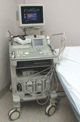
 Ultrasound waves are sound waves of a much higher frequency than we can detect - humans can hear up to around 20 kilohertz, but medical ultrasound operates at between 2 and 18 megahertz. In modern ultrasound machines, the sound waves enter the body from the probe containing an acoustic transducer, which produces the sound waves. These enter the body and bounce back off the internal organs in different ways depending on their density, coming back to the detector as an 'echo'. The time taken for the echo to travel back to the detector allows the depth of the object causing the reflection to be calculated. Solid objects bounce back a different signal to liquids, which is why it is easy for this technique to 'see' a foetus in the womb, full of amniotic fluid.
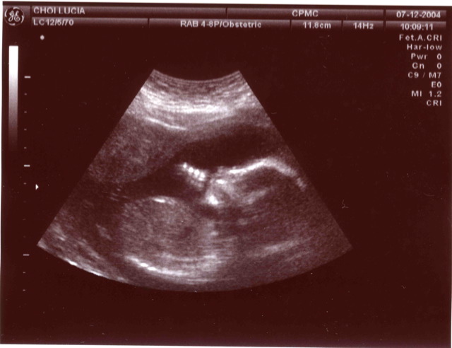
 Most people would recognise (or may have experienced) the use of sonography during pregnancy to image the baby, but it is also used as a diagnostic tool elsewhere in the body such as imaging organs like the bladder and prostate in men to check for abnormalities like tumours. Other forms of ultrasound can be used therapeutically, and in fact this was developed before ultrasound's imaging capabilities were realised, to clean teeth at the dentist, to treat cataracts and even used to treat benign and malignant tumours.
Professor Donald had served in the Second World War, where he gained knowledge of radar and sonar. He theorised that something similar to sonar could be used to look for abnormalities in the human body, and in 1955 visited the boilermakers Babcock and Wilcox to use their industrial ultrasound equipment usually used to check for flaws in sheets of metal. He took various sorts of cysts and tumours extracted from patients at his hospital to see if they could be differentiated from each other and from normal tissue like muscle (using a steak!). He had some limited success, but recognised the technique's potential, so collaborated with Tom Brown, a medical physicist, and John MacVicar, another obstetrician, to refine the technique. Their experimentation and results in examining abdominal 'masses' (like tumours and cysts) were what came to be eventually published in the Lancet paper. It was after this that Donald recognised the use of sonography in measuring foetal growth my measuring the dimensions of the head in utero.
 Within a decade of the publication of the paper, the use of ultrasound had moved into the mainstream and had already improved. As Donald himself said 'any new technique becomes more attractive if its clinical usefulness can be demonstrated without harm, indignity or discomfort to the patient', and given the lack of side effects and its obvious benefits, it quickly became an essential diagnostic tool.
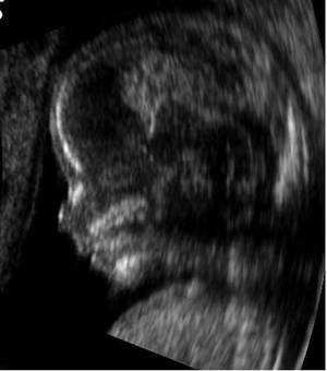
Ultrasonography is now used all the way through pregnancy, to check the growth and health of the baby and to determine the sex, as well as being important in tumour detection and diagnosis. It is easily portable and easy to use, as well as giving images in real time, so you can watch things like bloodflow and the heart beating, unlike other imaging techniques such as MRI that provide still pictures. There are limitations to the accuracy of ultrasonography, but it has no known long term effects and is an essential part of diagnostic medicine.
- Previous Hepatitis in the Clinic
- Next Printing Your Own House


Comments
Add a comment